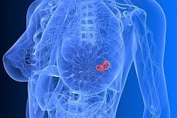
Medical imaging system comes above the radar
Bristol University spin-out Micrima (Bristol, UK) is developing a medical imaging system that can distinguish a breast tumor from normal tissue by detecting the difference between their dielectric properties.
The MARIA (Multistatic Array processing for Radiowave Image Acquisition) system, which does not expose patients to x-ray radiation, is expected to be better than conventional techniques at identifying breast cancer in younger women.
An ultrawideband radar pulse synthesized using a vector network analyzer sweeps in frequency from 4 to 10 GHz across the tissue. The signal is transmitted from each element in the multiple antenna array, after which it is received by all the other elements. A 3-D image of the breast is then reconstructed from the data that is received.
Because the transmitted radiowave signal has a peak power less than 1 mW, the public limits for exposure to radiowaves are not approached; therefore, the technology is intrinsically safe and is freely repeatable.
The technique was initially validated through computational modeling before it was experimentally validated using complex breast phantoms, or models of the breast using simulated tissues with the correct dielectric values for skin, fat, and tumors.
Micrima’s ultimate goal is to create a compact, low-cost version of the MARIA system that could be situated in surgeries and mobile screening units.
-- By Dave Wilson, Senior Editor, Vision Systems Design
