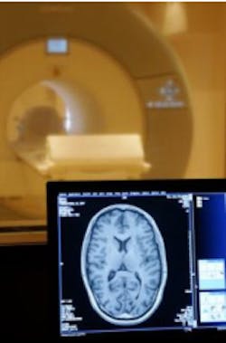
Imaging technique detects brain cancer
University of Oxford (Oxford, UK) researchers have developed a contrast agent that recognizes and sticks to a molecule called VCAM-1 that is present in large amounts on blood vessels associated with cancer that has spread to the brain from other parts of the body.
MRI scans show the distribution of the dye in the brain, enabling much smaller tumors to be detected than can be achieved using current techniques.
Cancer that has spread to the brain is extremely difficult to treat successfully. Small tumors can be treated with whole brain radiotherapy or surgery, and there are new chemotherapy treatments in development. But currently, it is only possible to detect larger secondary brain tumors, and these are more difficult to treat.
"We urgently need to find ways to diagnose these cancers at an earlier stage to improve survival rates. Our research suggests a new possible approach to do just this. The next stage is to build on these results and carry out clinical trials. If successful, we hope that early detection using this technique could increase the number of available treatment options for these patients," says Dr. Nicola Sibson of the Gray Institute for Radiation Oncology and Biology at the University of Oxford.
The research was funded by Cancer Research UK and the Wellcome Trust, with support from the National Institute for Health Research-funded Oxford Biomedical Research Centre.
-- by Dave Wilson, Senior Editor, Vision Systems Design