Smartphone system helps diagnose oral cancer
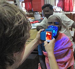
A Stanford University (Stanford, CA, USA) researcher has developed a way to use a smartphone to create detailed images of the oral cavity and screen patients’ mouths for suspicious lesions.
Assistant professor Manu Prakash oral cavity scanner -- called Oscan -- attaches to any smartphone's built-in camera, and allows an operator to take a high-resolution, panoramic image of a person's complete mouth cavity.
Illuminated by the device's blue fluorescent light, malignant cancer lesions are easily detected as dark spots. Images can then be sent wirelessly to health workers, dentists or oral surgeons for diagnosis, anywhere in the world. The device is designed for mass production, with an estimated material cost of just a few dollars.
The Oscan system, which is in the early phases of testing, has already been recognized with two awards from the Vodaphone Americas Foundation. This year, the OScan team won first place at the mHealth Alliance awards and came in second place at the Vodaphone Americas Foundation Wireless Innovation Project awards.
Dr. Prakash and the OScan development team plan to use the $250,000 in award money to perform more field testing of the device in India.
The OScan project was funded by the Wallace H. Coulter Foundation, which supports the translation of ideas that address unmet medical needs into treatments and devices that improve human health. Stanford University has filed for a patent on the technology.
-- Dave Wilson, Senior Editor, Vision Systems Design
