Imaging system to help diagnose psoriasis
A team from the Rochester Institute of Technology (Rochester, NY, USA) is seeking to improve the diagnosis and assessment of psoriasis through the creation of a multimodal, image-based analysis system.
"Presently, dermatologists use a method called the Psoriasis Area Severity Index (PASI) score to analyze affected areas of the skin," says Professor Christye Sisson, a biomedical photography at RIT and leader of the project team. "However, the process can be very subjective as each dermatologist could potentially grade patients differently."
The lack of empirical data can make it difficult to compare assessments made by different dermatologists or by different research groups studying the disease.
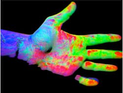
To resolve that issue, Sisson is working with Professor Francisco Tausk from the University of Rochester Medical Center to create an imaging tool that will be able to assess psoriasis based on the three criteria currently used by the PASI score: thickness, redness, and scale.
The team is currently testing several imaging techniques, including thermal, ultra violet reflectance, and LIDAR, to identify the best method for assessing each criteria. By doing so, Sisson and Tausk hope to ultimately create a standardized multimodal imaging system that could be implemented by dermatologists and hospitals nationwide.
"By modifying imaging technology that has already been developed we can create a repeatable, quantifiable method for diagnosing and ultimately treating patients with this disease," Sisson says.
Recent related medical imaging articles from Vision Systems Design that you might also be interested in reading.
1. Spectroscopy aids in characterizing skin cancers
A joint US-Dutch team under the direction of Anita Mahadevan-Jansen from Vanderbilt University (Nashville, TN, USA) has developed a noninvasive probe capable of both morphological and biochemical characterization of skin cancers.
2. Laser-based imager determines severity of burns
A laser-based imaging system developed by Aïmago (Lausanne, Switzerland) -- a startup in the science park at École Polytechnique Fédérale de Lausanne (EPFL; Lausanne, Switzerland) -- shows how blood circulates in bodily tissue.
3. Thermal imaging measures muscle soreness
Scientists from Loma Linda and Asuza Pacific Universities have employed thermal imaging to help in quantifying muscle soreness.
4. Hyperspectral imaging helps diagnose ulcers
Researchers at the UCLA Henry Samueli School of Engineering and Applied Science (Los Angeles, CA, USA) have developed algorithms that can analyze hyperspectral images of the foot to identify ulcers associated with diabetes weeks before they are visible to the eye.
5. Software helps classify skin cancer
Researchers at the University of Edinburgh (Edinburgh, UK) have developed diagnostic software that can help health care workers to correctly identify different types of skin lesions, leading to more effective diagnosis of skin cancer.
-- Dave Wilson, Senior Editor, Vision Systems Design