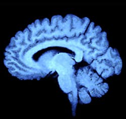
Spectroscopy aids brain tumor detection
UK researchers have shown that infrared and Raman spectroscopy - coupled with statistical analysis - can be used to differentiate between normal brain tissue and different tumor types.
Currently, when surgeons are operating to remove a brain tumor it can be difficult to spot where the tumor ends and normal tissue begins. But the team of researchers led by Professor Francis Martin at Lancaster University (Lancaster, UK) has shown it is possible to spot the difference between diseased and normal tissue using Raman spectroscopy, giving accurate results in seconds.
The development means it is now theoretically possible to test living tissue during surgery, helping doctors to remove the complete tumor whilst preserving intact adjacent healthy tissue.
The technique was also able to identify whether the tumors arose in the brain or whether they were secondary cancers arising from an unknown primary site. This could help reveal previously undetected cancer elsewhere in the body, improving patient outcomes.
"These are exciting developments which could lead to significant improvements for individual patients diagnosed with brain tumors. We and other research teams are now working towards developing a sensor which can be used during brain surgery to give surgeons precise information about the tumor and tissue type that they are operating on," says Professor Martin.
More information can be found here.
Recent articles on Raman spectroscopy from Vision Systems Design.
1. Spectroscopy detects explosives over long distances
Researchers at the Vienna University of Technology (Vienna, Austria) have developed a system to detect chemicals inside a container over a distance of more than a hundred meters.
2. Spectroscopy aids in characterizing skin cancers
A joint US-Dutch team under the direction of Anita Mahadevan-Jansen from Vanderbilt University (Nashville, TN, USA) has developed a noninvasive probe capable of both morphological and biochemical characterization of skin cancers.
3. Spectroscopy sniffs out fake spirits
Researchers at St. Andrew’s University (St. Andrews, Fife, Scotland) are using a microfluidic device coupled with a laser-based near-infrared (NIR) spectroscopy system to characterize whiskies and identify fakes.
-- Dave Wilson, Senior Editor, Vision Systems Design
