Cameras help image live biological specimens in 3-D
Researchers at laboratories in Europe and the US have created new microscopes capable of imaging rapid biological processes in thick samples that can be used to study live biological specimens.
Philipp Keller designed the microscope used by the group from the Howard Hughes Medical Institute (Ashburn, VA, USA), while Lars Hufnagel led the team from the European Molecular Biology Laboratory (EMBL; Heidelberg, Germany).
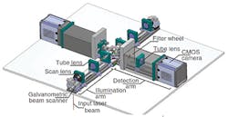
Called SiMView by Keller and Multi-View SPIM (MuVi-SPIM) by Hufnagel, the new 4-axis light-sheet microscopes build upon the SPIM (Selective-Plane Illumination Microscopy) technology developed by Keller and Stelzer at EMBL.
Like SPIM, the new microscopes shine a thin sheet of light on the sample, illuminating one layer at a time to obtain an image of the whole sample.
However, the SiMView and MuVi-SPIM microscopes use two diagonally-opposed light sources and two cameras to capture four images from different angles, eliminating the need to rotate the sample. This speeds up the imaging and enables the four images to be merged into a single high-quality, 3-D image for the first time.
Both teams independently chose the 5.5Mpixel Neo sCMOS camera from Andor (Belfast, UK) for their systems which offer frame rates of up to 100fps, and vacuum thermoelectric cooling to -40C to minimize dark current and maintain low noise.
More information can be found here.
Recent article on microscopy from Vision Systems Design that you might also be interested in.
1. Imaging technique shows nanoparticles do not penetrate skin
Research by scientists at Bath University (Bath, UK) is challenging claims that nanoparticles in medicated and cosmetic creams are able to transport and deliver active ingredients deep inside the skin.
2. Cell analyzer spots cancer in the blood
An automated optoelectronic image capture and processing system acquires images of cells in the blood and screens them for cancer in real time, with a record-low false positive rate.
3. Spectroscopy images Landau levels for the first time
Physicists have directly imaged Landau levels for the first time since they were theoretically conceived of by Nobel prize winner Lev Landau.
Vision Systems Design magazine and e-newsletter subscriptions are free to qualified professionals. To subscribe, please complete the form here.
-- Dave Wilson, Senior Editor, Vision Systems Design
