Low dose X-ray system aids breast disease diagnosis
Computed tomography (CT), an X-ray technique that allows a precise 3-D visualization of the organs in the human body, cannot be routinely used for the diagnosis of breast cancer because of the risk of the long-term effects of ionizing radiation.
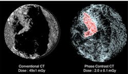
Recognizing these limitations, scientists have now developed a new technique that can produce 3-D computed tomography (CT) images with a spatial resolution 2-3 times higher than present hospital scanners, but with a radiation dose about 25 times lower.
The multidisciplinary team that developed the system comprised physicists, radiologists and mathematicians from the European Synchrotron Radiation Facility (ESRF, Grenoble, France), the Ludwig Maximilians University (Munich, Germany) and the University of California, Los Angeles (UCLA, Los Angeles, CA, USA).
The team combined three ingredients that may one day make CT scans for early detection of breast cancer possible -- high energy X-rays, a special detection method called "phase contrast imaging" and the use of a mathematical algorithm known as "Equally Sloped Tomography" (EST) to reconstruct CT images from X-ray data.
To demonstrate the effectiveness of the technique, the team X-rayed a human breast at multiple different angles using phase contrast tomography and applied the EST algorithm to 512 images to produce 3-D images of the organ.
In a blind evaluation, five independent radiologists from the LMU ranked the generated images as having the highest sharpness, contrast, and overall image quality of any of the techniques currently used to create 3-D images of breast tissue.
"This new technique can open up the doors to the clinical use of computed tomography in breast diagnosis, which would be a powerful tool to fight breast cancer," says Prof. Maximilian Reiser, Director of the Radiology Department at the LMU.
Today, the technology is in the research phase and will not be available to patients for some time. To be implemented in clinics, it needs an X-ray source small enough to be used for breast cancer screening. On a positive note, many research groups are now actively working to develop such a device.
Related articles from Vision Systems Design that you might also find of interest.
1. CT scan technique differentiates types of lung damage
A team from the University of Michigan Medical School (Ann Arbor, MI, USA) has used a technique called parametric response mapping (PRM) to analyze CT scans of the lungs of patients with chronic obstructive pulmonary disease (COPD).
2. Kinect could help medics estimate radiation dose from CT scanner
Researchers at the Hospital of the University of Pennsylvania are investigating whether a system based around the Microsoft Kinect could help predict the precise dose of ionizing radiation that patients require during a CT scan by providing an accurate estimate of whole-body volume.
3. Cell-CT scanner may help to diagnose cancer faster
An imaging system under investigation at the Biodesign Institute at Arizona State University (Tempe, AZ, USA) may help researchers pinpoint subtle aberrations in the nuclear structure of cells, leading to an improvement in the time it takes to diagnose cancer.
-- Dave Wilson, Senior Editor, Vision Systems Design
