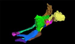
MIT system captures 3-D images of zebrafish larvae
Researchers at MIT (Cambridge, MA, USA) have built an automated system that can rapidly produce 3-D, micron-resolution images of thousands of zebrafish larvae and precisely analyze their physical traits.
According to MIT researcher Mehmet Fatih Yanik, zebrafish are genetically similar to humans and have many of the same developmental pathways. Hence the system can be used to provide a comprehensive view of how potential drugs affect vertebrates.
Using the new technology, researchers can grow larvae in tiny wells and flow them through a channel to an imaging platform. Once there, the embryos are rotated and 320 2-D images are taken from different angles, after which a 3-D reconstruction is performed.
Getting larvae to the platform takes about 15 seconds, and the imaging takes only 2.5 seconds. This allows hundreds or thousands of larvae to be imaged within hours.
The researchers also created a computer algorithm that can measure hundreds of traits and used that information to create a comprehensive phenotype map -- the overall description of an organism’s characteristics -- for each larva. This enables rapid and detailed studies of how different drugs affect those phenotypes.
This kind of analysis could be valuable for drug developers who need to efficiently screen thousands of drug candidates.
The software that the researchers wrote to generate the 3-D images is available on their website here.
The research was funded by the National Institutes of Health, the Packard Award in Science and Engineering, a Sparc Grant from the Broad Institute, and a Howard Hughes Medical Institute Student Fellowship.
Related articles from Vision Systems Design that you might also be interested in.
1. Fruit flies tracked by vision
Using multiple synchronized and calibrated cameras, researchers in the UK and the US have built a vision system that enables them to detect and keep track of the behavior of fruit flies.
2. Vision system assesses the gait of pigs
Newcastle University (Newcastle, UK) researchers are using a video motion capture system to assess what constitutes a 'normal' gait in pigs by focusing on the angle of their joints and length of stride.
3. High-speed cameras capture moth motion
Tyson Hedrick, an assistant professor in the Department of Biology at the University of North Carolina at Chapel Hill (Chapel Hill, NC, USA) is using three high-speed cameras to determine what makes hawk moths so skilled at flying.
-- Dave Wilson, Senior Editor, Vision Systems Design
