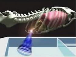
One of the methods used to treat a tumor is to focus ultrasound waves to heat cancer cells to 60C - destroying them and leaving healthy tissue largely unharmed.
But to use the technique to treat liver tumors presents a major problem because the organ shifts back and forth during breathing. This increases the risk that the ultrasound beam path misses the cancer cells and instead heats the surrounding healthy tissue too strongly.
For this reason, researchers have only applied this method to patients under general anesthesia. To treat a tumor with ultrasound, a medical ventilator is paused for a few seconds so that the patient remains absolutely still.
To solve this problem, European researchers working on the so-called FUSIMO (Focused UltraSound In Moving Organs) project are developing a system that tracks the movement of the liver during surgery using MRI data and then applies the beam of ultrasound at the appropriate time to treat tumors in it.
In principle, the procedure could be applied to other abdominal organs that move with breathing and which are difficult to target with the ultrasound beam path such as the stomach, kidneys, and duodenum.
The FUSIMO project began in 2011 and has been funded for three years with 4.7m Euros. Eleven institutions from nine countries are involved. The project is coordinated by the Fraunhofer Institute for Medical Image Computing MEVIS (Bremen, Germany).
More details can be found on the FUSIMO web site here.
Related items from Vision Systems Design that you might also find of interest.
1. Ultrasound images from a pill
A group of researchers from Dundee University (Dundee, Scotland, UK) and several other research institutions have been awarded £5m in grant funding from the Engineering and Physical Sciences Research Council (EPSRC, Swindon, UK), to develop a new system that will transmit ultrasound images from a pill.
2. Software enhances ultrasound images
An Oxford University (Oxford, UK) spin-out has developed software that enhances the detail and field of view of conventional ultrasound images.
3. Analyzing ultrasound helps diagnose cancerous tissue
An assistant professor of physics at Kettering University (Flint, MI, USA) is seeking a better way to diagnose between chronic pancreatitis and pancreatic cancer by analyzing endoscopic ultrasound images.
4. 3-D ultrasound software company spins out of Oxford
University of Oxford (Oxford, UK) spin-out Intelligent Ultrasound (Oxford, UK) has raised £610,000 to develop software that can reduce the risk of incorrect or missed diagnoses from ultrasound scans and avoid costly, inconvenient rescans.
-- Dave Wilson, Senior Editor, Vision Systems Design


