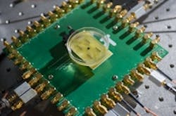
CMOS image sensor to provide real-time 3D images from inside the heart and blood vessels
Researchers from the Georgia Institute of Technology have developed a catheter-based device equipped with a CMOS image sensor that would provide forward-looking, real-time 3D images from inside the heart, coronary arteries, and peripheral blood vessels, which would help better guide surgeons during heart procedures.
The single-chip device integrates an ultrasound transducer array with a front-end CMOS image sensor to provide 3D intravascular ultrasound and intracardiac echography images. The dual-ring array, which is on a single 1.5 mm silicon chip, includes 56 ultrasound transmit elements and 48 receive elements. In the center of the device is a 430 µm hole to accommodate a guide wire. In addition, the array is able to operate with just 20 mW of power due to power-saving circuitry that shuts down sensors when they are not needed, which reduces the amount of heat generated inside the body.
On-chip processing of signals allows data from more these elements to be transmitted using just 13 cables, allowing it to travel through blood vessels. Images produced by the device would provide significantly more information than existing cross-sectional ultrasound, according to a press release.
A prototype developed by the researchers at the institute was able to provide image data at 60 fps, which is an encouraging sign for its future adoption in surgical procedures, according to F. Levent Degertekin, a professor in the George W. Woodruff School of Mechanical Engineering.
"Our device will allow doctors to see the whole volume that is in front of them within a blood vessel," he said in the press release. "This will give cardiologists the equivalent of a flashlight so they can see blockages ahead of them in occluded arteries. It has the potential for reducing the amount of surgery that must be done to clear these vessels."
The team expects to conduct animal trials to demonstrate the device’s potential applications and ultimately expect to license the technology to an established medical diagnostic firm and conduct the clinical trials necessary to obtain FDA approval.
View the press release.
Also check out:
Wearable glasses enable surgeons to visualize cancerous cells
Robot vision system picks stem cells
Chemical imaging technique could improve diseased tissue analysis
Share your vision-related news by contactingJames Carroll,Senior Web Editor, Vision Systems Design
To receive news like this in your inbox, click here.
Join our LinkedIn group | Like us on Facebook | Follow us on Twitter | Check us out on Google +
About the Author

James Carroll
Former VSD Editor James Carroll joined the team 2013. Carroll covered machine vision and imaging from numerous angles, including application stories, industry news, market updates, and new products. In addition to writing and editing articles, Carroll managed the Innovators Awards program and webcasts.
