GigE cameras provide vision in cell-imaging microscope system
When developing its NyONE cell analysis microscopy system, SynenTec required the use of two GigE cameras, one which would be used to acquire microscopic images of a cell, and one that would monitor the optimal position of an object in relation to the sample holder.
SynenTec’s NyONE device is used for automated cell analysis primarily in cell research and drug discovery applications. The device provides a detailed look into the interior of cells and helps scientists understand the process within the cell while mapping its tracks digitally and converting them into data. To do so, image processing and digital cameras are a necessary part of the system. SynenTec chose two Baser ace GigE cameras, one that acts as the microscope’s main camera, and the other that monitors an object’s positioning.
Basler ace cameras are available in VGA to 14 MPixel models with frame rates all the way up to 340 fps. The cameras have a broad sensor portfolio, including CMOS, CCD, and CCD with NIR-enhancement versions, from Sony, CMOSIS, and Aptina. The 29 mm x 29 mm cameras have a number of interface options, including USB 3.0, GigE, and Camera Link. Based on the camera description for the NyONE device, it seems likely that the specific model used was Basler’s acA2040-90um, which is a 2048 x 2048 pixel monochrome USB 3.0 camera with a 5.5 µm x 5.5 µm pixel size that achieves a frame rate of 90 fps.
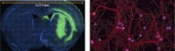
With the NyONE system, tissue or blood samples are placed on a sample holder and strained with fluorescent dyes that bind specifically to the cell or the tissue to facilitate the examination of special areas in or on the cell via images captured by the ace camera.
"Image-based measuring instruments are becoming increasingly important for the life sciences," said Matthias Pirsch, managing director at SynenTec in a news release. "The quality of Basler cameras is at a level that allows us to replace alternative measuring methods with high-quality imaging systems."
He added, "The pylon driver architecture also permits us to make application-specific adjustments quickly and inexpensively and thereby react to market changes rapidly."
View the case study.
Also check out:
Medical Imaging: Polarization subtraction system characterizes cancer
Embryoscope imaging device may improve chances of In Vitro Fertilization
Researchers developing terahertz detectors with eye on improving certain imaging applications
Share your vision-related news by contacting James Carroll, Senior Web Editor, Vision Systems Design
To receive news like this in your inbox, click here.
Join our LinkedIn group | Like us on Facebook | Follow us on Twitter | Check us out on Google +
About the Author

James Carroll
Former VSD Editor James Carroll joined the team 2013. Carroll covered machine vision and imaging from numerous angles, including application stories, industry news, market updates, and new products. In addition to writing and editing articles, Carroll managed the Innovators Awards program and webcasts.