Smart microscope aids diagnosis
Computer-aided imaging system automatically quantifies cell characteristics for pathologists.
By David Lieberman, Contributing Editor
Vision systems are helping automate the life sciences in numerous, and potentially critical, ways. Pathology labs and associated medical operations, for example, can benefit from using a semi-automated microscopy system. Such a system could aid in clinical management of cancer, including detection, classification, and treatment.
To accomplish this, TriPath Imaging and Ventana Medical Systems have developed the Ventana Image Analysis System (VIAS), which is an interactive, computer-aided microscopy system designed to help pathologists understand diseased cells. It does this by capturing images from an immunohistochemically stained slide and providing a quantitative analysis of the cells on that slide. As the image of a particular region of interest (ROI) is captured, the system simultaneously captures its x-y coordinates. Equipped with this information and a motorized stage, the microscope can repeatably return to any previous ROI the pathologist selects.
AmeriPath, a provider of diagnostic health-care information, has been using the VIAS. According to Scott Sittler, director of anatomic pathology at the company’s Center for Advanced Diagnostics in Orlando, FL, USA, the system provides more accurate results in certain cases, including “small-volume tumors and tumors that have a particular growth pattern that makes them more difficult to evaluate.”
LOOKING AT CELLS
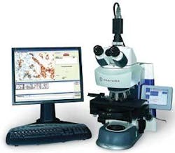
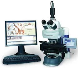
The system is based on a semi-automated microscope equipped with a digital camera, encoded turret, and motorized stage. The VIAS also includes a PC with a frame-grabber board running TriPath’s proprietary image-analysis software. The peripherals include a 19-in.-diagonal LCD monitor, a wireless mouse and keyboard, a barcode reader, and a smart card reader. The microscope is equipped with a small monochrome LCD with a touch-sensitive overlay for user input (see Fig. 1).
The digital microscope integrated into the VIAS is a custom version of the Axio Imager.M1 from Carl Zeiss Micro Imaging, equipped with a motorized stage from Märzhäuser Wetzlar. It uses a 100-W halogen bulb for illumination and contains five objective lenses on its turret, providing 2.5X, 5X, 10X, 20X, or 40X magnification. The turret is encoded to ensure that all ROI images in a particular sequence of images are captured with the same objective. The system can provide quantitative results for images captured at either 10X or 20X magnification.
The Zeiss microscope has its own memory, which stores the setup parameters used with each objective. Items such as light setting, aperture, and focal point are displayed on the small LCD.
A CAMERA EMBEDDED
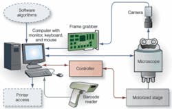
The color CCD camera embedded in the microscope is the Model IK-TF5 from the Imaging Systems Division of Toshiba America Information Systems. The three-chip, progressive-scan camera measures 1.73 × 1.73 × 3.07 in. and weighs 5.74 oz. It has a high-precision prism block assembly that provides a signal-to-noise ratio (S/N) of 62 dB, allowing the camera to deliver color fidelity with minimal illumination. The Zeiss microscope communicates with the PC over a USB 2.0 link; the camera, in turn, uses an analog composite-video link to communicate with the frame grabber (see Fig. 2).
“We selected the IK-TF5 for overall image quality and performance,” says Tim Macri, senior director of systems engineering at TriPath. Macri says that progressive scan was an essential image-capture requirement for the VIAS. “Progressive scan allows image acquisition while the microscope stage is still moving and minimizes the effects of vibration, which is a major issue for interactive imaging systems.”
Based on three 1/3-in. CCDs, the IK-TF5 contains square 7.4 × 7.4-µm pixels arranged in a 659 × 494-pixel format. Horizontal scan takes place at 31.469 kHz and vertical scan at 59.94 Hz. The camera’s electronic shutter operates between 1/100 and 1/100,000 s, and the maximum frame rate is 60 frames/s at full resolution. In partial-scan mode, the camera accommodates up to 180 frames/s. It provides selectable gain from -3 to +18 dB and a choice between automatic white balance or a manual selection in the range between 3200K and 5600K.
PROCESSING THE IMAGE
The PC is a custom VIAS-optimized configuration from Pen-Tech running Windows XP and containing a Intel Pentium 4 microprocessor, a minimum 512 Mbytes of system memory, and a 160-Gbyte hard drive. In addition there are three PCI expansion slots and interfaces including RS-232 serial, parallel, and USB. An RS-232 port is used for the interface between the PC and a discrete stage-control box, which interfaces to the motorized stage over a proprietary Märzhäuser electromechanical interface. One VIAS configuration also contains a 100 BaseT Ethernet port for connection to Ventana’s Benchmark family of slide staining systems.
One of the three expansion slots is populated by a Meteor-II/Multi-Channel frame-grabber from Matrox Imaging that interfaces to the Toshiba camera by means of an analog video interface and a trigger signal. The frame grabber has real-time image transfer to memory for live video in a window at sustained rates of up to 130 Mbytes/s. An on-board 4-Mbyte data buffer, in turn, ensures reliable image capture even under high bus latency conditions, which typically occur in systems with concurrent image capture, display, graphics, network access, disk access, and general external I/O.
The VIAS comes equipped with a wireless USB keyboard and mouse, a 19-in. color ViewSonic LCD monitor, a smart card reader, and a USB barcode reader. The barcode reader, Model 9540 Voyager from Metrologic Instruments, is for scanning in demographic information from a slide stained by a Benchmark system. The reader is capable of decoding all standard 1D, RSS-14, RSS Limited, and RSS Expanded barcodes.
Barcoding a slide “cuts down on a lab technician’s time and the requirements for setting the instrument up,” reports AmeriPath’s Sittler. Moreover, “as long as the information on the barcode was originally entered correctly, this reduces the likelihood of switching specimens because of a labeling error,” he adds.
Ventana places the VIAS with an end user for free, while charging a per-slide usage fee. The system is also equipped with an Advanced Card Systems Model ACR38U smart card reader connected to the PC via USB. The reader accepts a smart card that serves as a debit card for processing slides.
PUTTING THINGS TOGETHER
The system is designed to fit into the traditional work flow of a typical pathology operation. However, the pathologist can capture images and view them on a large color monitor, which also displays a thumbnail gallery of a sequence of captured images. Once an image is captured, the system provides a quantitative assessment.
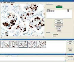
Selecting an ROI image from the gallery for review or comparison automatically moves the objective lens to the same location on the slide where the actual ROI resides (see Fig. 3). This allows the pathologist to further explore the ROI using different settings for magnification, focus, light intensity, and aperture.
The system measures “how much positivity the tissue is expressing for certain markers, which will affect the treatment protocol,” explains Ventana’s Jon Minken. In other words, it measures what percentage of cells react to an antibody, as visually indicated by the optical density of a stain that binds to that antibody, based on a predefined threshold. By reporting the percentage of marker-expressing cells in an ROI, the system helps the pathologist interpret the tissue and determine the best therapy.
To aid the pathologist in interpreting a case of breast cancer, for example, if the cells on a slide demonstrate a certain amount of positivity of expression of estrogen receptor, then that patient is a candidate for Tomoxifin or other hormonal therapy, Minken says. Likewise, a positive score when measuring expression of the HER-2/neu gene qualifies a patient for Herceptin.
GETTING STARTED
The pathologist begins using the VIAS by selecting a test type from a list in the application software. The most common types include ER (estrogen receptor), PR (progesterone receptor), and HER-2/neu. The pathologist then opens a new case and starts inspecting a slide in the normal fashion. Meanwhile, a live image of the slide is displayed on an approximate 8 × 8-in. window on the 14.8 × 11.9-in. LCD screen at a 1:1 pixel ratio.
The VIAS software, which runs under Windows, contains image-processing algorithms developed by TriPath, including the Chromogen Separation method and multisegmentation engines. Chromogen Separation is a transformation from the RGB color space of the camera to the color characteristics of the dyes used for each of the available VIAS test types. The transformation defines the spectral fingerprint of each dye in terms of RGB contribution, resulting in a higher S/N for the image processing that follows.
The multisegmentation engine architecture, in turn, optimizes the distinction between cells and background for each class of test. A further optimization takes place through the use of Slide Type and Panel data structures that contain the specific parameters for each test type on the system.
Using the VIAS, objective lenses are changed in the conventional manner. The pathologist turns the microscope turret. Typically, a slide is scanned with a 2.5X or 5X objective lens, and the stage is moved manually to inspect different areas of the slide in search of ROIs, and then focused in at 10X or 20X magnification. The VIAS provides an auto-selection feature, which automatically moves the stage to display the most likely tumor area. However, the pathologist can override this and take manual control at any time.
The system includes a set of simple drawing tools for the pathologist to modify these areas as desired. The circle and square icons on the screen establish boundaries for ROI selection within an area of a slide, and a pencil tool defines an irregularly shaped area. However, when the user decides to capture an image, a click of the wireless mouse selects the Grab button on the LCD. The system automatically provides a quantitative report on the positivity of the image.
null