Imaging system automates memory recall analysis
To judge the effectiveness of drugs developed to improve memory loss in patients with diseases such as Alzheimer’s, researchers at Maastricht University (Maastricht, the Netherlands;www.psychology.unimaas.nl) have developed an imaging system that is being used to judge memory recall in rats. Developed by Huub Hamers of the Faculty of Psychology and Neuroscience and Jeffrey Habets of VITechnologies (Weert, the Netherlands; www.vi-tech.nl), the system is designed to analyze the effects of the drugs and how they can improve memory recall.
“In 1988,” says Hamers, “it was discovered that while object recognition is performed visually by human beings, mammals such as rats perform the same function by sniffing the object.” To observe the nature of this memory recall, psychologists and engineers from Maastricht designed a closed object arena in which a number of different objects were placed. After the rat is placed into this object arena for a period of 3 minutes, the rat sniffs the objects and the time the rat’s nose is less than 2 cm from the object is scored. After this period, the rat is withdrawn from the arena and some of the objects replaced with new objects. One hour later, the rat is placed back into the arena and the process repeated.
“If the rat remembers the features of objects,” says Hamers, “it will spend more time sniffing the new objects. If this repeated test is performed approximately 24 hours after the original test, however, the rat will have no recall about the location or features of the objects.” Because measuring the time the rat’s nose is within a specific distance of these objects allows a comparative measurement of memory recall to be made, the memory recall of rats given medication can be compared to control animals that have not been given such medication. In this manner, the effectiveness of these drugs can be observed.
“In the past,” says Hamers, “scoring the time the rat spent close to a specific object was a task performed manually and was subject to the observer’s judgment. Using the automated system, this is no longer the case.” To accomplish this automated measurement, a monochrome analog camera was placed approximately 250 cm from the field of view of the arena. Images from this camera were then digitized into a PC using a PCI-1411 analog frame grabber from National Instruments (NI; Austin, TX, USA;www.ni.com).
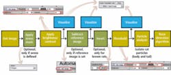
As images are captured, a number of different algorithms are used to compute both the position of the nose of the rat and the duration of the rat’s nose from the object (see Fig. 1). Developed using NI LabVIEW and the NI Vision Development Module, an arena mask is first applied to localize the region of interest. After adjusting brightness and contrast to ensure that the objects are visible, a reference image of the arena is subtracted to eliminate the image background.

In this way, only the objects within the arena are visible to the imaging system. “In some cases, brown as opposed to white rats are used in the experiments,” says Hamers. “Because of this, an optional contrast inversion algorithm can be applied that will allow a brown rat to be imaged.” Thresholding this image then allows the rat to be isolated from the scene as it moves around the arena.
To judge the exact position of the rat’s nose, however, required some quite ingenious thinking on behalf of the researchers. In developing their “nose detection” algorithm, the researchers realized that the center of gravity (COG) of the rat must first be computed. Then, by iteratively computing the line of maximum intercept crossing the center of mass, the exact position of the rat’s nose could be determined (see Fig. 2).
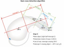
Before this could be accomplished, however, it was necessary to segment the tail from the body of the rat. Although this task could have been accomplished using image-processing techniques, a far easier solution was found. By highlighting the base of the tail of the white rat with a black marker, image segmentation techniques were used to isolate the tail, prior to computing the position of the rat’s COG and nose.
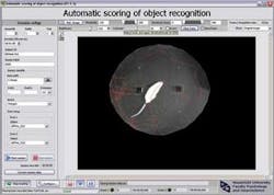
After the animal was introduced into the arena, the position of its nose was then automatically tracked and displayed graphically on a PC along with the duration spent near the object (see Fig. 3). According to Hamers, the system has already found great interest among European drug companies wishing to test the effects of new memory recall drugs such as Donepzil.
null