X-ray Imaging Delivers a Better Cut

Winn Hardin, Contributing Editor
In New Zealand, 24 million lambs are processed for export every year. A typical boning room will process up to 600 lambs per hour, which involves reducing the animal from dressed carcasses down to boxes of specified cuts. This is a repetitive, labor-intensive task involving dozens of people and dangerous machines such as rotary knives.
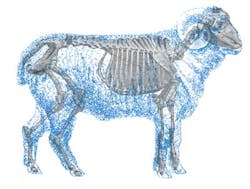
Scott Technology, a system integrator that specializes in the design and manufacture of automated production and process machinery, has automated the lamb boning task. The system developed by Scott Technology uses an x-ray imaging system, a combination of commercially available and custom image-processing software, and a programmable logic controller (PLC) to direct automated rotary knives to separate the carcass into the correct portions based on the internal skeletal structure of the animal. The system also helps eliminate safety concerns, alleviate the pressure of finding seasonal workers, provide consistency in cutting, and increase yield (see Fig. 1).
Initial cuts
The first processing task is to take the full carcass and cut it into three primal pieces—the forequarter, middle, and hind legs. The forequarter is separated by a planar cut between the ribs and must provide a specified number of ribs in the forequarter, or leave a specified number of ribs in the middle. Each carcass will have a different height and pitch angle for the cut. The middle can be separated in various ways, but all involve a horizontal cut at a height based on the location of the aitch bone (pelvis) of the carcass.
Attempts to accurately locate the position of bones using external features have not resulted in high enough accuracy. Even compared against the sophisticated vision-processing capability of workers manually using rotary knives, this process can be improved. Scott Technology chose to use x-rays to directly image the bones and provide unambiguous positioning data.
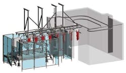
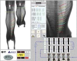
The carcasses enter the room suspended by their hind legs from a chain conveyor. To accommodate a new carcass every 6 s, the chain drive passes between an NDI-225-FB-Be (Fan Beam) x-ray tube from Varian, which is driven by a Gulmay high-voltage generator, and a Sens-Tech x-ray detector. Scott Technology uses collimators (lead shields) to reduce the 90 × 30° fan from the Varian x-ray tubes to a more useful 60° planar fan shape (see Fig. 2).
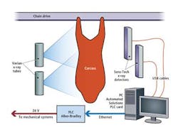
Sens-Tech offers a range of x-ray detector lengths, resolutions, and sensing characteristics. Scott Technology chose a 2048-mm-long detector with 1.6-mm pitch and a total resolution of 1280 pixels. The 16-bit output allows sufficient contrast to be present in dense regions such as the shoulders and more diaphanous regions such as the rib cage, while the 1.6-mm pitch provides sufficient spatial resolution to achieve the required rotary-knife cut accuracies. The detector plugs straight into a PC, running MVTec Software’s Halcon 8.0 via a USB connection.
The end result of the imaging system is a pair of 16-bit grayscale images of each carcass. The carcass passes twice between the x-ray tube and detector to yield two images taken from different angles to allow the software to generate a 3-D map of the lamb’s skeleton, using triangulation based on data from the two x-ray shadowgraphs.
Biological variability
Initially the images are segmented with filtering, dynamic thresholding, blob analysis, and prior knowledge of the basic structure of a lamb carcass. Specific regions are further filtered, enhanced, transformed, and traced. Halcon’s shape-matching functionality is useful for locating features even though the system is dealing with greatly variable biological structures. The functionality provides quick localization, which enables simple refinements to get an accurate position.
One of the main challenges in analyzing lamb carcass images is determining an accurate rib count. Most lamb carcasses have 13 pairs of ribs, but a significant number of them have 12 or 14 pairs. Some even have differing numbers on each side of the spine. To achieve the specified cut position, all ribs must be detected—one cannot assume a total and count from whichever end is convenient. This becomes particularly difficult in the forequarter end of the carcass, where the thin first rib is among the forelegs, shoulder blade, and associated heavy muscles.
Blob analysis, edge detection, and shape matching techniques all fail to find this rib with the required accuracy. A clearer image of the first rib region cannot be obtained and still keep up with the rate of two images of an entire carcass every 6 s. However, Halcon allows several types of supervised learning methods to make classifications.
Scott Technology chose Halcon’s support vector machine (SVM) as the main method for teaching the machine-vision software how to locate the critical ribs and structures for cutting. The position of a potential first rib is extrapolated from the other ribs, which appear with greater contrast in the x-rays and are easier for the vision system to identify.
Various features in the region of interest where the first rib should be located are extracted, such as positions, shapes, and edge intensities. The region of interest for the first rib is concentrated in a vector of 100 numbers that represent critical features. The SVM takes this feature vector and returns a classification: rib or not a rib.
This determination is based on the results from manually training the system on thousands of carcass examples and allowing the system to “learn” how to detect a first rib’s position. The SVM splits the 100 dimensional feature space into regions, each representing a single class. When a new unseen feature vector arrives, the SVM simply sees where it lies in the feature space and returns which region or class that is. Given the nature of the biological product, cases near the border between regions can occasionally be misclassified, yet the system has achieved 98% accuracy in obtaining the correct rib count.
All possible cuts
Once the specified rib has been found on both images, its edge is traced. This pair of 2-D curves from the two different viewpoints can then be combined to give the 3-D curve of the rib. A best-fit plane then determines the cut position. All possible cuts—six different forequarter separation cuts and four different middle separation cuts—are calculated for every carcass (see Fig. 3).
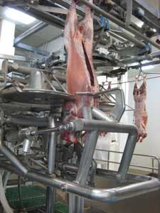
The data are fed through to the Allen-Bradley PLC via Ethernet connection. The PLC controls the various blades and mechanical systems. All the mechanical equipment is designed and built by Scott Technology, including stabilization belts to keep the carcass steady while scanning, and servo-pneumatic systems to transfer the product smoothly and position the cutting blades.
A single PC controls the x-ray detector, x-ray controller, image analysis, a user interface (see Fig. 4), communications with the PLC, and logging and archiving of images. The vision PC maintains its own list of images. These are matched with a trigger and associated ID number from the PLC. When the cut positions for a carcass are calculated, the information is simply sent to the PLC along with the matching ID. The PLC program manages all the tracking of product and data.
The operator can select different cuts from an Allen-Bradley RSView touchscreen. The speed of the image analysis, in combination with a modest multicore processor, is easily able to keep up with the required throughput.
The x-ray imaging system and primal cutting machine is in operation at several lamb-processing plants around New Zealand. More than 2 million lamb carcasses have been imaged and processed. Manual inspection of the primal pieces coming off the end of the machine has shown the system provides a consistent accuracy greater than 98%.
Company Info
Allen-Bradley
Milwaukee, WI, USA
Automated Solutions
Santa Rosa, CA, USA
MVTec Software
Munich, Germany
Scott Technology
Dunedin, New Zealand
Varian Medical Systems
Palo Alto, CA, USA
Vision Systems Articles Archives