Simple fingerprint casts made from materials such as clay or gelatin can fool most fingerprint recognition devices. In fact, a recent study funded by the U.S. Department of Homeland Security found that wearing fake prints, known as "spoofing" in law enforcement circles, can beat a standard fingerprint scanner 80% of the time. This is because traditional fingerprint scanning devices use optical systems that only image the very top layer of skin. Along with being tricked by spoofing, these devices sometimes fail to recognize legitimate prints when the finger being scanned is dirty, worn, scarred, or too wet or dry.
To overcome this, Egidijus Auksorius at the Langevin Institute (Paris, France; www.institut-langevin.espci.fr) has developed a fingerprint imaging system that permits the visualization of internal skin layers from which the external fingerprint regrows. Not only are these internal layers less affected by the damages sustained on the surface, they are also less susceptible to spoofing.

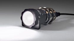
The internal fingerprint sensor is based on a variation of a full-field optical coherence tomography (FF-OCT) technology, known as oblique FF-OCT (o-FF-OCT). In the design of the system, a 150W halogen bulb is coupled to a 5mm diameter fiber bundle. The output of this bundled light is then imaged through an aperture diaphragm. A secondary lens then images this aperture to infinity. This light is then divided into both reference and sample beams by a beam splitter.
To image the internal characteristics of a fingerprint, a finger is pressed against a 5mm flat glass window. Light returning from the reference and sample beams then creates an interference pattern that is imaged by a solid-state camera. To image all the layers under the skin, a reflector is placed in the reference path that is tilted. The angular tilt of the reflector defines the depth span of o-FF-OCT image in a specimen and can be chosen so that it contains details of the external fingerprints, sweat pores and the internal fingerprints in one image. This can be used to determine the depth of the internal fingerprints (or sweat ducts). The final image of, for instance, the internal fingerprints can then be recorded by re-tilting back the reflector to the normal orientation so that an en face image would be recorded.

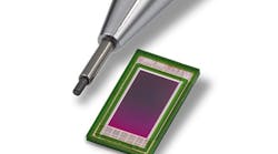
Initially, Auksorius and Boccara chose a Xeva 640 InGaAs camera from Xenics (Leuven, Belgium; www.xenics.com) to acquire 640 x 512 images at 25 fps in the infrared part of the light. Recently, however, this camera has been replaced with a Q-2A750 CoaXPress (CXP) camera from Adimec (Eindhoven, The Netherlands; www.adimec.com) that operates in the visible spectrum and uses a 2MPixel global shutter image sensor to deliver images of 1440 × 1440 pixels at 720 fps.

To transfer captured image data to the PC, the Q-2A750 camera is interfaced to a PCI Express-based Cyton CXP4 CoaXPress frame grabber from BitFlow (Woburn, MA, USA; www.bitflow.com). Using this camera/computer interface combination, images of an internal fingerprint can be captured in under a second, which is adequate for live fingerprint imaging applications. Speed is important because any finger movement will compromise the quality of the image.
As well as being applied in biometric applications, Auksorius and Boccara predict that the o-FF-OCT system they have developed may also be used to study and monitor various skin diseases and conditions due to its ability to image deep under skin.