Integration Insights: Embedded machine vision system detects liquid level
Smart camera advances improve liquid level detection for medical applications
Robert Geiger
Used in clinical diagnostics to verify the volume of reagents or samples in containers, liquid-level detection facilitates the automation of various tests and helps to ensure the accuracy of results. Many clinical systems in the field today use capacitive, ultrasonic, pressure and even weight to determine the level or volume of a liquid.
While these methods can be very effective in certain applications, each may have limitations depending on the application needs and requirements. For example, weighing a sample requires the use of an individual scale or load cells for each sample. While effective, this method is generally too cumbersome for the high-volume, high-throughput systems seen in commercial applications such as medical and clinical chemistry.
Level measurement methods
Other approaches to liquid-level detection include capacitive, ultrasonic and pressure all of which require some type of contact with the container such as probes or other system equipment, which tends to slow down the process and increase time to result. Such methods require that the sensor have an open path to the top of the liquid. In the case of a test tube or ampule, for example, the top would have to be removed or pierced which can lead to contamination of the sample and even breakage of system equipment.
Ultrasound methods require that the cap be removed so that sound waves can be measured off of the surface of the liquid. This opens up the opportunity for spillage during handling and potential contamination with the surface of the liquid. While the cap can remain on the container for pressure methods, it must be pierced so that a volume of air can be injected and measured to calculate the liquid level. Piercing the cap also increases the possibility for contamination of the sample.
Moreover, these methods require one probe per sample. So either there are many probes with their own cables, mechanical mounts and power, which adds to system complexity and costs, or the samples have to be presented to the probe one at a time, which can reduce system throughput. Capacitive sensors are yet another technique used for liquid level detection, but are known to be less accurate than other methods.
Due to recent advances in embedded smart cameras and image processing technology, machine vision has become an effective technique regardless of container type. For example, embedded machine vision and image analysis are used to detect liquid level in test tubes, cuvettes, pipette tips and ampoules, and offer engineers designing clinical chemistry and medical diagnostic equipment many advantages over tactile methods.
Advantages of machine vision for liquid-level detection
Of the potential methods mentioned above, embedded medical machine vision is emerging as a front-running technology for determining liquid level. As previously mentioned, no technique is without limitations, but machine vision is fast becoming recognized as a uniquely versatile, non-invasive and efficient approach.
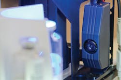
With a camera-based system, an image of the sample(s) is taken and processed (Figure 1). Therefore, there is no need to remove or puncture the top of the container or come in con tact with any part of the container at all. This ensures the purity of the sample and eliminates contamination or breakage of tubes, probes or other system equipment.
Also, due to the camera-based nature of a machine vision system, it can be utilized to determine the level of multiple samples at one time. Multiple containers can be imaged in a single field of view and processed accordingly. In fact, advanced methods are being developed that will enable level detection from above for microtiter trays or other multiple-compartment containers.
Other advantages of a machine vision system include the ability to determine volume based on the liquid level of different types of containers. Capacitive or ultrasound techniques will only give the lab technician information about the top of the liquid relative to a known point. Volume has to be inferred from assumptions on the type of container used.
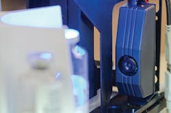
If using different types of test tubes, for instance 75mm and 100mm test tubes, the level could be measured but the volume would have to be calculated based on a previous knowledge of the type of tube used. With a vision-based system, the liquid level can be measured and the tube type could be determined via image processing to give an accurate volume determination on the fly (Figure 2). Imaging can also simultaneously determine the container size in order to give a true volume measurement.
In addition, image-based systems afford users the ability to determine layers within the sample. This can be very useful in the determination of the separate layers or blood fractionation that occurs after centrifuging to distinguish plasma, buffy coat and red blood cells.
In some cases, a biological sample would need to be agitated as part of the process. This can result in a liquid sample that contains a large amount of bubbles or foam on the top of the sample. Embedded machine vision systems can be engineered to distinguish these occurrences from the coherent liquid layer and, if required, report that back to the system for further analysis.
A picture is worth a thousand words
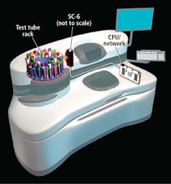
Undoubtedly, the most unique and advantageous aspect of a vision-based system is that it takes a picture of the sample being analyzed. As labs strive to automate for increased throughput and efficiency, no other liquid level measuring technique offers the versatility of image capture and analysis (Figure 3). Beyond basic liquid level and volume, machine vision and image analysis can provide lab technicians with even more details about a given sample in real-time, including:
• Meniscus shape to give an indication of the characteristics of the sample
• Color of the sample or individual layers
• Illumination intensity to determine reaction process over time
• Verification and tracking by 1D/2D barcodes
• Container morphology and volume calculations
• Images can be saved for archival purposes
Conclusion
There are many ways to determine the liquid level and volume of samples being analyzed in a clinical device. Careful thought should be given to the efficiency, complexity and throughput considerations of the system, as well as the users' needs and requirements. Like many available techniques, embedded medical machine vision is a proven method for determining liquid level (Figure 3). However, unlike other methods, image processing provides an unmatched level of flexibility and versatility, leading to increased throughput and reduced errors
Robert Geiger, Senior OEM Systems Engineer, Jadak, North Syracuse, NY, USA (www.jadaktech.com)


