Imaging system aids in heart diagnosis
Advanced medical-imaging system captures, processes, and stores detailed artery images for local or remote access and viewing.
By David Head
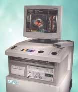
Medical-imaging OEMs such as JOMED Inc. (Rancho Cordova, CA, USA) are developing products for minimally invasive vascular intervention, as well as developing intravascular ultrasound (IVUS) imaging systems and diagnostic imaging catheters. For example, the company's In-Vision Gold IVUS system incorporates image-acquisition, transfer, and display technologies. This system comprises a custom imaging engine, a PC-based graphical user interface (GUI), and a data-storage and retrieval system.
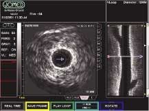
Doctors at interventional laboratories, such as cardiocatheter labs, use IVUS catheter to determine the types and sizes of lesions in patients' coronary arteries. The catheter is inserted into the femoral artery, through the heart, and into the coronary arteries where lesions are imaged from the inside out using a tiny beam-forming transducer that sends and receives images. The transducer generates sound waves that reflect differences in body tissue densities, and the echoes returning from these reflections are compiled into vectors. The vectors represent specific tissue data that, when combined with other vectors, can produce a visual display of the area reflecting the transducer sound waves.
Different parameters are added to or subtracted from this information to compensate for image attenuation, gain, brightness, and contrast. The vector information is then compiled into a format that can be sent to a display monitor. This array-based technology provides more precise details than those obtained by standard x-ray imaging. It also alleviates nonuniform rotational distortion, so doctors can more accurately identify and diagnose the magnitude of an occlusion.
"A key challenge we faced during the years of design and development of the In-Vision Gold system was miniaturization. Our goal was to make the components extremely small while giving the system the ability to capture images in real time," says Craig Lindsay, JOMED global senior product manager, imaging. The company's research-and-development efforts in this area and the availability of ever-smaller components have resulted in a threefold reduction in the size of the imaging system compared to the original version released ten years ago, making it faster, more portable, and easier to use.
Image generation
The In-Vision image generation starts with the imaging catheter. The catheter's 64 transducer elements are mounted around its circumference to produce a full 360° cross-sectional image of the patient's blood vessel. This catheter connects to a patient interface module (PIM), which excites the catheter's transducer elements to transmit ultrasonic energy to the surrounding tissues. The transmitter/receiver echo information is brought through the PIM to the system's custom imaging engine.
This imaging engine comprises seven custom and off-the-shelf boards: analog-to digital, beam-forming, reconstruction, color flow, digital vector processing, scan converter, and VME/PCI bridge. The analog-to-digital converter board converts the analog echo data to digital data. Processing of the digital data is controlled by the reconstruction board, which coordinates the accumulation of echo data for processing in the beam-forming board. The reconstruction board also coordinates catheter control through the PIM.
From the beam-forming board, echo data are compiled by the digital vector processing board, where vector data are mapped and formed vector by vector to produce an entire display. The data can then be formatted by the scan-converter board into a raster-data information format. The scan-converter board is connected to the VME/PCI bridge board, which transfers data processed by the imaging engine into the system's 866-MHz Pentium III-based PC. Last, the data are passed through the CPU/CPI bridge board to the system monitor.
Eight still images and up to nine minutes of video can be viewed on a fluoroscopy monitor. These images can be saved to a hard drive, archived to CD-ROM or videotape, or transferred to a remote server via DICOM (Digital Imaging and Communications in Medicine). This networking standard allows information exchange between medical devices and the transfer of image and patient information to a DICOM system workstation or server over the Internet using DICOM Standard 3.0.
System operation
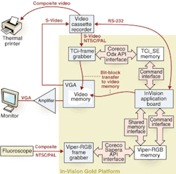
The In-Vision Gold imaging system interfaces to a medical fluoroscope in either a high- or low-resolution composite NTSC/PAL format (see Fig. 1). A Coreco Imaging (St.-Laurent, QC, Canada) Viper-RGB frame grabber synchronizes and digitizes the input signals at either 8 or 10 bits. This single-slot, color and monochrome video-acquisition and processing board for the PCI bus features 40-MHz acquisition and can acquire and transfer images in real time to system memory. It provides RGB acquisition, three 10-bit ADCs, and a signal-to-noise ratio of 55 dB. When PCI bus traffic is heavier than normal, the frame grabber buffers image data into local memory temporarily so that processing speeds are not affected.
"We chose the Viper-RGB frame grabber because it enables real-time video output of 30 frames/s. Medical personnel can display patient information directly on a fluoroscopy monitor or store the data in digital format so that they can review it at any time on their PC monitors," says Lindsay.
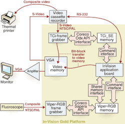
The frame-grabber data are transferred via a Coreco Sapera API software interface to another Viper-RGB frame grabber and then on to an In-Vision application board. The Sapera software library offers more than 300 image-acquisition, processing, and analysis functions. The application board transfers the fluoroscopic data via Sapera's DirectDraw software to the system's video memory and 17-in. monitor (see Fig. 2).
While data are displayed on the monitor, a doctor can overlay and combine other GUI attributes with the IVUS and fluoroscope video images so that all relevant data can be compared and displayed side-by-side. Once displayed, VGA images can be sent via an S-Video output to an external VCR for archival. Recorder functionality is controlled by the In-Vision application board over an RS-232 serial link. When archival data are needed, a Coreco 24-bit color TCi-SE frame grabber (now replaced by the company's Bandit-II series) digitizes and transfers image data from the VCR directly to video memory for display. At this time, a doctor can draw overlays on top of the video for further diagnostic inputs.
The digitized VCR signal displayed on the PC's VGA monitor, gives doctors the option of VCR playback (see Fig. 3). This board offers pixel-packing capability that supports multiple data-output formats.
Coreco's Oculus-TCI software enables direct-draw capabilities and image transfers. "In addition to providing quality video, there are a range of image viewing, storage, and retrieval options to suit the needs of each doctor," Lindsay comments. "Integrating all of the components in the In-Vision Gold system results in an affordable feature set."
For doctors, the system provides clear and reliable images from which to make diagnoses, an easy-to-use interface, and flexibility to view images at whatever time and in whatever format they prefer. For patients, it means more accurate diagnoses, shorter exam times, and less discomfort.
DAVID HEAD is applications engineer, Coreco Imaging, St.-Laurent, QC, Canada; www.imaging.com.
Info
Coreco Imaging www.imaging.com
JOMED Inc. www.jomed.com


