Thermography aids chiropractic diagnosis

Vision system couples IR imager with blackbody temperature references to evaluate blood circulation.
By Lawrence J. Curran, Contributing Editor
An English company and a university clinic in Wales are collaborating in a project using infrared thermography that holds promise for diagnosing various neurovascular pathologies, including Raynaud's disease. This disease interrupts the passage of blood to body parts such as a finger, by causing a spasm of the small terminal branches of an artery or arterioles.
Researchers at the Welsh Institute of Chiropractic at Glamorgan University are using a Land Instruments International FTI Mv thermal imager as the central sensor in a PC-based system. Laboratory tests indicate that the system can be a diagnostic tool, with the potential to help clinical specialists make an objective assessment of the underlying condition.
Land Instruments has developed a method of linking the camera and its software to two precise blackbody temperature reference sources, which have been certified for accuracy by the UK National Physical Laboratory. The resulting thermal data can be rendered as a series of color images that can be used to differentiate normal and abnormal changes in temperature. The portable, moderately priced apparatus offers the advantage over competitive equipment of having the blackbody references closely coupled to—and providing temperature calibration for—the camera. The company has applied for a patent and is preparing to market the system.

In an image of a man's hands taken one minute before a cold-provocation test, red hues indicate higher IR radiation, which relates to blood flow (left). Immediately after a cold-provocation test, during which the hands were immersed in cold water for one minute, blood flow is drastically reduced as indicated by the lower levels of IR radiation (middle). Ten minutes after the cold-provocation test, the fingers have not rewarmed completely (right). Subject has a condition similar to Raynaud's disease, in which blood flow in the terminal ends of digital blood vessels has an exaggerated response to cold stimulation.
A. I. "Drew" Heusch and P. W. McCarthy at the institute are using the system. Among other neurovascular phenomena under investigation, the feasibility of measuring the response of a cold-provocation test on subjects who have experienced episodes of vasospasm in their hands during winter is being explored. Vasospasm is the sudden constriction of a blood vessel, causing a reduction in blood flow to the affected area.
Although the research project is at an early stage, the Welsh team considers the system's ease of use, portability, and inclusion of the temperature references to be of great advantage in a clinical situation. Subjects for testing are referred by chiropractors at the institute. Some of the subjects have complained about tingling and other sensations in their fingers after exposure to cold outdoor temperatures.
During testing, ambient-temperature images of a subject's hands are taken at intervals of 0, 1, 5, and 6 minutes. If they show no cooling effect, they undergo a cold-provocation test, in which the subject's hands are placed in plastic gloves, then immersed in cold water for one minute. The gloves are then removed, and additional images are taken at 8, 13, 18, 23, and 28 minutes. A plot of the changes at various times in temperature difference (distal-proximal) for each digit—including those taken at ambient—are a good indicator of how quickly normal blood flow returns to the fingers after cold provocation.
The protocols used are standardized. The subjects must have had no manipulative therapy or shower within at least the previous four hours, wear no rings or shirts with tight cuffs, and eat only a light meal before the test. The room used for testing must also be environmentally controlled.
PARTIONING THE SYSTEM
The test system relies on the FTI Mv thermal imaging camera, along with the two blackbody reference sources, which were designed and developed by Land Instruments (see figure on p. 50). The camera incorporates an uncooled microbolometer focal-plane array of 160 × 120 pixels.
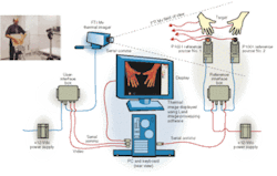
The reference sources are located in the camera's field of view. The camera is connected to the system's Pentium II-based PC through a universal interface box (UIB) over an RS-422 serial communications link. The blackbody references also connect to the PC through a reference interface box (RIB), using another RS-422 link. The camera has its own power supply, as do the blackbody temperature references.
A MuTech Mv510 frame grabber and an Amplicon dual-port serial communications card areside on the PC. Land's LIPS Mini software is used in processing the data for presentation as thermal images, which can help diagnose abnormalities.
Ian Ridley, product specialist for the design and development of imaging products at Land Instruments, says the thermal-imaging camera measures the temperature of a subject's hands by collecting the infrared radiation from them. "The system achieves very high accuracy by having the camera collect the infrared from the two blackbody references simultaneously with that from the hands to effectively calibrate each image."
Ridley will not say precisely how the references are implemented, except that they incorporate a microcontroller, which monitors and controls the temperature of their aluminum housing and enclosure. They are heated and cooled by a thermoelectric (Peltier) heater.
Permanently positioned in the field of view of the imager, these calibrated reference sources have their temperatures set by the thermographer to accommodate the subject and type of test to be conducted, Ridley says. They provide a reference area in the live image scene. The imager is then adjusted to maintain this reference area at a fixed radiance value (200). The subject's hands are placed between the reference sources, at approximately 1 m from the camera.
IMAGE PROCESSING
After the image has been acquired, it is analyzed by Land's image-processing software, which displays the images and records them to disk. The two reference temperature values are transmitted by the RIB to the PC. The reference values in the area of the image are used to calibrate the image temperatures.
Land's LIPS Mini software provides five false color palettes, which are applied to the images of the subject's hands, indicating IR radiation from the hand that relates to blood flow. The subject whose hand images are shown on p. 49 has a condition with symptoms similar to Raynaud's disease.
Before acquiring the Land Instruments system, this kind of testing was possible using research equipment, according to Heusch, but the IR imaging and temperature calculations had to be done separately. Further, the temperatures obtained from the IR images had to be manually modified to take into account the traceable standard blackbody temperature, which was taken simultaneously.
With the current setup, "we know exactly what the temperatures are, because once in place, the blackbody references give us two traceable standards that accurately calibrate the camera via the software," says Heusch. He adds that the system is small, lightweight, easily portable, and easy to store, making it attractive for use in a chiropractic clinic. "We wanted a camera that we could recommend for our clinical chiropractors to help them assess if their treatments are working."
Additional testing is being performed to qualify the system for routine clinical use. McCarthy says that, in addition to the hand studies already under way, the clinic anticipates using the system to help diagnose and track treatment efficacy for shoulder and back conditions, as well.
Company Info
Amplicon Liveline, Brighton, UK www.amplicon.co.uk
Land Instruments International, Dronfield, UK www.landinst.com
MuTech, Billerica, MA, USA www.mutech.com
UK National Physical Laboratory, Teddington, UK www.npl.co.uk
Welsh Institute of Chiropractic at Glamorgan University, Pontypridd, UK www.glam.ac.uk