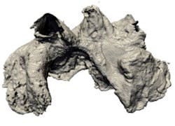
Researchers at the School of Physics and Astronomy at Manchester University (Manchester, UK) have built a computational model of an anatomically correct sheep's heart.
It was made by taking a series of very thin slices of the heart, imaging them in 2-D and then using a computer program to render them into a 3-D model.
The reconstruction includes details of the complex fiber structure of the tissue, and the segmentation of the upper chambers of the heart into known distinctive atrial regions. Single-cell models that take into account information about the electrical activity in different atrial parts of regions the heart were then incorporated into the model.
Professor Henggui Zhang created the model to investigate the medical condition known as atrial fibrillation (AF).
Professor Zhang and his team focused on the pulmonary vein which is a common area that initially triggers AF. They simulated erratic electrical waves passing through the vein and the surrounding atrial tissue, and then studied the impact this had on the rest of the heart.
They discovered that regional differences in the electrical activity across the tissue of the heart -- known as electrical heterogeneity -- is key to the initiation of AF. The largest electrical difference was between the pulmonary vein and the left atrium which may go some way to explaining why the pulmonary vein region is a common source of irregular heartbeats.
The scientists also identified that the fiber structure of the heart plays an important role in the development of AF. There were directional variations in the conduction of electrical waves along and across the fibers known as anisotropy. The fiber structure in the left atrium is much more organized compared with the complex structures of the pulmonary vein region. The sudden variation in conduction at the junction between the left atrium and the pulmonary vein regions appeared to contribute to the development of AF.
"We have identified the individual role of electrical heterogeneity and fiber structure in the initiation and development of AF. It has not previously been possible to study the contribution of the two separately but using our computational model we've been able to clearly see that both electrical heterogeneity and fiber structure need to be taken into consideration when treatment strategies for AF are being devised," says Professor Zhang.
Recent articles on 3-D imaging that you might also find of interest.
1. 3-D modeling improves spinal casting process
Engineers at 4DDynamics have developed a 3-D system that can be used to develop prosthetic corsets in less than half a day.
2. 3-D vision optimizes robotic car parking
serva transport systems and in-situ Vision & Sensor Systems have developed a fully automated car parking system that makes use of more of the space in a parking garage by first identifying a vehicle and then transporting it to an empty parking slot using a self-guided robot trolley.
3. Robots team with 3-D scanners for fast part profiling
AV&R Vision & Robotics (Montreal, QC, Canada) has developed a system that combines visual inspection, robotics, and 3-D profiling techniques.
Vision Systems Design magazine and e-newsletter subscriptions are free to qualified professionals. To subscribe, please complete the form here.
-- Dave Wilson, Senior Editor, Vision Systems Design


