How robots could remove blood clots with medical imaging and steerable needles
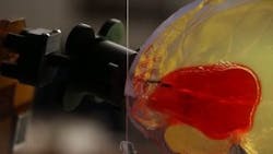
A robot-based surgical system under development at Vanderbilt University uses steerable needles and medical imaging technology to penetrate the brain with minimal damage and remove deadly blood clots.
For the past four years, assistant professor Robert J. Webster III and his team had been working on a steerable needle system for transnasal surgery, according to a Vanderbilt press release. Specifically, Webster was focusing on operations that involve the removal tumors in the pituitary gland and at the skull base that traditionally involve cutting large openings in the skull or face. So when Webster attended a conference in Italy and heard Marc Simard, a neurosurgeon at the University of Maryland School of Medicine, talk about how useful a needle-sized robot arm that reaches into the brain to remove clots would be for physicians, he knew his system would be right for the job.
The system—which Webster calls an active cannula—consists of thin, nested tubes that each has a different intrinsic curvature. By rotating, extending, and retracting the tubes, an operator can steer the tip in different directions, enabling it to follow a curving path. The system, which is much simpler than the previous transnasal concept, works by first determining the location of a blood clot with a CT scan. Once the position is determined, the surgeon picks the best point on the skull and the proper insertion angle for the probe. The angle is dialed into a fixture called a trajectory stem, which is attached to the skull immediately above a small hole that has been drilled.
Next, the surgeon positions the robot so it can insert a straight outer tube through the trajectory stem and into the brain. The surgeon then selects the smaller inner tube with the curvature that best fits the shape of the clot, and attaches a suction pump to the external end piece and places it in the tube.
Using CT scan imaging for guidance, the robot then inserts the outer tube into the brain until it nearly reaches the surface of the blood clot, then inserts the inner tube into the clot and the pump is turned on, which sucks out the blood clot. The robot rotates, extends, and retracts the tubes to remove the clot. The team reports that the robot was able to remove up to 92% of simulated blood clots in feasibility studies.
One part that is still being worked out, according to Webster, is after the removal of the majority of the clot when external pressure can cause the edges of the clot to partially collapse, which makes it difficult to track the clot’s boundaries. In future developments, the team hopes to add ultrasound imaging combined with a computer model of how brain tissue deforms to ensure that all of the clot material can be removed safely and effectively.
View the Vanderbilt press release.
Also check out:
Image processing software increases the visibility of catheters
Robot uses infrared camera, lighting to draw blood
3D imaging machine helps physicians identify cancer earlier, more frequently
Share your vision-related news by contacting James Carroll, Senior Web Editor, Vision Systems Design
To receive news like this in your inbox, click here.
About the Author

James Carroll
Former VSD Editor James Carroll joined the team 2013. Carroll covered machine vision and imaging from numerous angles, including application stories, industry news, market updates, and new products. In addition to writing and editing articles, Carroll managed the Innovators Awards program and webcasts.
