One of the numerous technologies and applications being highlighted at Photonics West 2014 was a non-invasive optical imaging system used to quickly obtain images at the cellular level to assist in diagnoses and biopsies.
CAReIOCA began in 2012 as a collaborative project funded by the European Commission’s Seventh Framework Programme. The project saw the development of an imaging system with the purpose of providing a medical tool for better localization and analysis of cancer biopsies in the operating room in order to "reduce failure and reoperation rates in pre-operative diagnosis." CAReIOCA’s technical partners, LLTech, CMOSIS, and Adimec collaborated on the technical aspects of the imaging system.
First, the group laid out four work objects for the development of the system:
- Development of a CMOS image sensor designed for full-field optical coherence tomography (FFOCT). Specifications for a custom sensor have been defined by CMOSIS and the newly designed sensor is expected to multiply the speed of FFOCT imaging by 20X. Sensor prototyping is currently underway, with initial results expected in March.
- Development of a CMOS camera for FFOCT imaging. Specifications of the camera have been set by Adimec in tandem with CMOSIS and LLTech. Adimec is designing the camera architecture with a focus on power efficiency and FPGA performance. The camera, which will be CoaXPress, is expected to produce images by June.
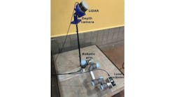
- Production of two clinical prototypes. Two research instruments have been mounted and delivered to the clinical partners, Leiden University Medical Center and the Gustave Roussy Cancer Campus, for an early definition of reading and diagnosis criteria. Clinical requirements have been set, and the design of the instrument is currently underway, with special attention on optical architecture, miniaturization, and safety procedure compliance. The first distal images using the instrument have already been obtained.
- Clinical evaluation. Clinical protocols have been defined, including definition and number of samples, clinical procedure, and regulatory requirements. The first FFOCT images were obtained with the idea of producing an image library that helps to determine reading criteria for FFOCT images and diagnosis methods.
CAReIOCA was developed and built with the idea of integrating the “perfect” camera to an FFOCT device, which would assist in diagnoses, particularly of cancer, as well as the quality control of biopsies. In addition, such FFOCT devices may eventually find use in other applications, including robotic surgery, endoscopic resections, or in-situ treatment at microscopic scales, according CAREeIOCA.
View more information on CAReIOCA.
View a blog post by Adimec on Photonics West.
Also check out:
Polyp-detecting pill camera receives FDA clearance
Chemical imaging technique could improve diseased tissue analysis
Imaging software tracks tumor rotation during radiotherapy
Share your vision-related news by contactingJames Carroll, Senior Web Editor, Vision Systems Design
To receive news like this in your inbox, click here.
Join our LinkedIn group | Like us on Facebook | Follow us on Twitter | Check us out on Google +