Medical Imaging: Robot performs IV procedures autonomously
Venipuncture-the puncture of a vein-is a medical procedure most commonly used to withdraw blood. Performed over 1.4 billion times each year in the United States, the success of the procedure varies significantly depending on the patient's physiology and the practitioner's experience. Because of this, a number of systems have recently been introduced that use commercial imaging devices to enhance the visibility of veins.
One such system is the AccuVein AV400, a hand-held vein illumination system from AccuVein (Cold Spring Harbor, NY, USA;www.accuvein.com). In operation, reflected infrared light from the skin's surface is digitized, processed and re-projected onto the skin providing an image that maximizes the contrast between the veins and other tissue.
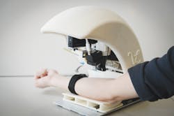
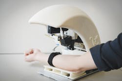
While useful, such devices still require a skilled clinician to perform the needle insertion. Now, Dr. Alvin Chen and his colleagues at VascuLogic (Piscataway, NJ, USA;www.vasculogic.com) have developed a portable, image-guided, medical robot prototype that automates this procedure. According to Chen, the VenousPro system improves the accuracy and safety of the venipuncture procedure with close to 100% accuracy.
Taking less than two minutes to perform the procedure, the VenousPro system images and maps the 3D spatial coordinates of forearm veins to robotically direct a needle into the designated vein. To control the operation of the system, a cRIO-9025 800MHz real-time embedded controller from National Instruments (NI; Austin, TX, USA;www.ni.com) is interfaced through an Ethernet hub to two Flea3 GigE cameras with IR sensitivity from Point Grey (Richmond, BC, Canada; www.ptgrey.com).
These cameras are configured as a stereo pair and use standard camera calibration, rectification, and stereo correspondence algorithms from NI's Vision toolbox combined with custom software. The cameras are used to image the veins and to track the needle before it is placed into the vein. Once positioned, data from an ultrasound probe is digitized through the USB port of the cRIO system to track the needle.
"The 3D NIR system provides a coarse, "zoomed-out" map of all of the veins in the patient's forearm and calculates the 3D coordinates of the selected vein relative to the robot," explains Chen. "The ultrasound probe is then lowered over that location to provide zoomed-in high-resolution images of the selected vein. The ability to measure blood flow (to detect collapsed veins) and track the needle as it enters the tissue are important added benefits of ultrasound," says Chen.
The miniature robot used to position the needle is comprised of a three-degree-of-freedom (DOF) XYZ gantry system and a four (XYZ and rotation) DOF robot arm. The XYZ gantry uses linear stages to position the robot arm, the cameras and the ultrasound probe to the correct position. The four DOF robot arm is then used to position the needle. All four DOF are required to align the needle at the proper orientation, move along a nonlinear trajectory if required, and then to insert the needle.
"Separating the mechanics in this way allows the robot arm to be miniaturized by minimizing the torque requirements at the distal end of the arm-the needle is very lightweight, whereas everything else is heavier," says Chen.
To control the miniature robot used to position the needle, four NI 9514 C Series servo drive modules are used to direct the four degrees of freedom required. Bi-directional communication to the robot is then performed using a single NI 9401 C Series digital I/O module. Also central to the CompactRIO system are three linear stages that connect to the RS232 port to provide XYZ positioning.
To correlate joint angles in the robotic arm with the 3D Cartesian coordinates of the needle tip, kinematics, proportional-integral-derivative controller (PID), and path-planning virtual instruments (VIs) in the NI LabVIEW Robotics Module were used.
To predict future position and velocity of the next frame in real-time, NI's LabVIEW Control Design and Simulation Module was used. Here, a Kalman filter is used to predict the "true" needle tip position and velocity based on the feedback from the imaging systems and the readout from the encoders of each motor in the robot arm. "Since there will be some random variation associated with both imaging and encoder systems, the Kalman filter is a powerful way to account for this and allow all of the data to be used to control the needle," says Chen.
To date, VascuLogic has demonstrated more than 98% accuracy in multiple in-vitro studies using the system. Although at present, the CompactRIO system requires an external PC to display the images generated by the system, VascuLogic plans to upgrade to a new multicore CompactRIO hardware that will allow a direct VGA interface to a flat-panel monitor, eliminating the need for a host PC. Once these final design changes are implemented, the company will commence with Institutional Review Board-approved human clinical testing in preparation for an initial FDA filing.
Vision Systems Articles Archives