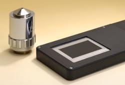
Fraunhofer IOF develops handheld, ultrathin microscope to catch melanoma
Researchers from the Fraunhofer Institute for Applied Optics and Precision Engineering (IOF; Jena, Germany), have created a very small, handheld microscope that can capture high-quality images in less than one second. To obtain such images of a relatively broad area, doctors or researchers typically use a microscope that scans the area in a grid pattern, recording many images one point at a time, then merging the images to form one complete picture. Among other applications, it could be used to examine suspect skin blemishes or perhaps check document authenticity.
The new microscope's imaging system consists of three glass plates, stacked one on top of the the other. Each plate is covered with a matrix of the tiny lenses on both top and bottom surfaces. Looking down through the plates from above, each lens lines up both with its counterpart on the other side of its plate, and with the other lenses that occupy the same location on the other plates. Microscopic details are therefore imaged through a stack of six lenses, along with two achromatic lenses. These stacks of lenses are called channels, and it is the images produced by the multiple channels that are digitally joined together, side-to-side and top-to-bottom, to image an area of 36 x 24 mm².
SOURCE: Fraunhofer IOF
--Posted by Vision Systems Design
