Vision-based blood test system delivers results in 15 minutes
Engineers at Biosurfit (Lisbon, Portugal) have developed a compact desktop blood analyzer called the “spinit” that could be used in doctors' surgeries.
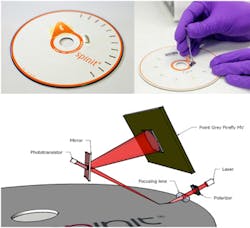
Using a combination of Point Grey (Richmond, BC, Canada) cameras, proprietary software, and DVD-style test cartridges, the spinit takes one small drop of blood, analyzes it and gives precise results within 15 minutes.
The CD-like cartridge in the spinit contains several detection zones that include a biological recognition layer (BRL) which contains antibody fragments immobilized on a gold surface that acts as a selective trap for a specific blood biomarker.
When placed in the spinit reader, the cartridge spins to a predetermined velocity and the centrifugal force forces the blood sample through the microfluidic structures of the cartridge.
When a patient's blood passes through the BRL, biomarkers bind to immobilized antibody fragments. To determine the concentration of biomarkers, the spinit uses Surface Plasmon Resonance (SPR), a well established spectrometry technique that enables molecular changes on the cartridge's biological recognition layer to be detected.
In use, the system measures the refractive index of light reflecting off the gold surface of the cartridge. The light source used to illuminate the surface of the cartridge comprises a near IR wavelength laser, a polarizer and a focusing lens. The optical detector uses a Point Grey Firefly MV FireWire monochrome camera acquiring images at almost 160FPS with an ROI of 50 x 800 pixels.
The camera is triggered when a phototransistor behind a semi-opaque mirror detects a reflection coming from the detection zone on the cartridge. The camera then acquires 3,000 images of each detection zone over a 10-minute period.
When the biomarkers bind to the immobilized antibody fragments, they cause a measurable refractive index change in the SPR signal which is measured by the Firefly camera. This change in the SPR is then used to determine the concentration of biomarkers in the blood sample.
The spinit will be released later in 2012 and the first cartridge will measure C-reactive Protein (CRP). CRP is an acute phase protein actively circulating in the body during an inflammatory response.
Recent articles on the use of vision systems in medical applications that you might also find of interest.
1. Medical imaging system comes above the radar
Bristol University spin-out Micrima (Bristol, UK) is developing a medical imaging system that can distinguish a breast tumor from normal tissue by detecting the difference between their dielectric properties.
2. Imaging combination delivers high-contrast, high-resolution images
Scientists from the University of Southern California (Los Angeles, CA, USA) and Washington University (St. Louis, MO, USA) have developed a new type of medical imaging system that gives doctors a new look at live internal organs.
3. Training system provides a better hand wash
Engineers at Glanta (Dublin, Ireland) have developed a mobile vision-based system that can train medical staff to improve their hand hygiene in healthcare centers.
4. Researchers develop software to image tissue in 3-D
Computer scientists and medical researchers at the University of Leeds (Leeds, UK) have developed a way of studying tissue samples using a digital scanning system that produces high-resolution, multi-colored 3-D images that can be rotated and examined from any angle.
5. Image sensor goes down the throat
An image sensor from OmniVision Technologies (Santa Clara, CA, USA) has been designed into Ambu's (Glen Burnie, MD, USA) aScope 2, a single-use intubation videoscope intended for medical procedures such as percutaneous dilational tracheostomy (PDT).
6. Imaging system sees bacteria behind the eardrum
A new medical imaging device invented by a research team led by University of Illinois (Champaign, IL, USA) electrical and computer engineering professor Stephen Boppart allows doctors to see biofilms behind the eardrum to better diagnose and treat chronic ear infections.
7. Imaging through the bowel backwards
An innovative scope developed by Avantis Medical Systems (Sunnyvale, CA, USA) provides a retrograde view or "rear view" to allow a physician to see behind folds in the colon wall, where cancers may be hiding.
-- Dave Wilson, Senior Editor, Vision Systems Design