Researchers image a hand with protons
Researchers developing a new medical imaging technology that uses protons instead of x-rays presented the first proton radiographic image of a hand last month at a medical imaging conference in Southern California.
The interdisciplinary team working on the project includes physicists and students at the University of California, Santa Cruz (Santa Cruz, CA, USA), medical researchers and doctors at Loma Linda University (Loma Linda, CA, USA), and computer scientists at California State University (San Bernardino, CA, USA)
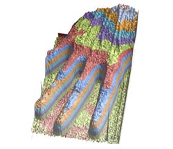
To create the image, the imaging team took a radiographic hand phantom (an anatomical model of a hand with the same radiographic properties as a real hand) which they imaged with protons from the medical proton synchrotron at Loma Linda University Medical Center (LLUMC).
"This image demonstrates the new promise of proton imaging, which is now within reach of becoming a new, potentially low-dose medical imaging modality," says Hartmut Sadrozinski, a research physicist at the Santa Cruz Institute for Particle Physics (SCIPP) at UC Santa Cruz. "Our ultimate goal is to do proton computed tomography and reconstruct the images in three dimensions, like an x-ray CT scan."
The proton image of the hand phantom clearly shows details of the bone structure. Unlike x-ray images, however, it also shows the soft tissue of the hand in detail.
The team presented their findings at the 2012 IEEE Nuclear Science Symposium and Medical Imaging Conference in Anaheim in October.
The research is supported by the National Institute of Biomedical Imaging and Bioengineering, the US Department of Defense Prostate Cancer Research Program and the United States-Israel Binational Science Foundation.
More information can be found here.
Recent articles on medical imaging that you might also find of interest.
1. Medical imaging system comes above the radar
Bristol University spin-out Micrima (Bristol, UK) is developing a medical imaging system that can distinguish a breast tumor from normal tissue by detecting the difference between their dielectric properties.
2. MRI system images muscles in 3-D
Researchers at the Technische Universiteit Eindhoven (TU/e; Eindhoven, The Netherlands) and the Academic Medical Center (AMC) in Amsterdam have developed a technique that allows muscle structures to be imaged in 3-D.
3. Researchers develop software to image tissue in 3-D
Computer scientists and medical researchers at the University of Leeds (Leeds, UK) have developed a way of studying tissue samples using a digital scanning system that produces high-resolution, multi-colored 3-D images that can be rotated and examined from any angle.
-- Dave Wilson, Senior Editor, Vision Systems Design