UK researchers to shrink MRI system
A new system that enables medical personnel to image difficult areas of the body is being developed by researchers at Imperial College London (London, UK).
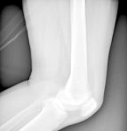
The team is making a compact Magnetic Resonance Imaging (MRI) system for hospitals that could improve the way that joints including knees and elbows are scanned. The system could help doctors to make more informed decisions about surgery, which could improve outcomes and recovery times for patients.
The team says that the finished MRI instrument will be the size of a coffee table, fitting neatly around the knee. Standard hospital MRI machines are currently the size of a small car and cost around £900,000 ($1.4m) -- a significant portion of which pays for installation. The new machines will be cheaper to manufacture, costing only an estimated £200,000 ($320,000).
Dr. Mihailo Ristic, from Imperial College's department of mechanical engineering, will be developing the new system which will involve the design of a new magnet that will be unlike the conventional horseshoe shaped or cylindrical magnets that are used in today's MRI systems.The magnet will be moveable and the field it produces will be able to be oriented at various angles to the patient's body.
The first year of the project, which runs for three years, will be spent perfecting the magnet design, using software developed by Ristic's research group. By the end of the project, the Imperial scientists aim to collaborate with industrial partners to produce a commercial product.
The system will be designed specifically to image knees, but the approach could also be used to image a wide variety of body parts.
Related articles from Vision Systems Design that you might also be interested in.
1. Algorithm cuts down MRI scan time
The time taken to perform a magnetic-resonance-imaging (MRI) scan could be cut from 45 minutes down to just 15 minutes, thanks to a new algorithm developed at the Massachusetts Institute of Technology (MIT; Cambridge, MA, USA).
2. MRI system images muscles in 3-D
Researchers at the Technische Universiteit Eindhoven (TU/e; Eindhoven, The Netherlands) and the Academic Medical Center (AMC) in Amsterdam have developed a technique that allows muscle structures to be imaged in 3-D.
3. New MRI-based technique looks inside solid objects
Researchers at Yale University (New Haven, CT, USA) have developed a new way of seeing inside solid objects, including animal bones and tissues, potentially opening a vast array of dense materials to a new type of detailed internal inspection.
-- Dave Wilson, Senior Editor, Vision Systems Design