X-ray source gets shrunk
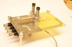
AUniversity of Missouri (MU; Columbia, MO, USA) engineering team has invented a compact system the size of a stick of gum that can be used to generate a source of x-rays.
"Currently, x-ray machines are huge and require tremendous amounts of electricity," says Scott Kovaleski, associate professor of electrical and computer engineering at MU. "In approximately three years, we could have a prototype hand-held x-ray scanner using our invention. The cell-phone-sized device could improve medical services in remote and impoverished regions and reduce health care expenses everywhere."
The system uses a mass of crystalline piezoelectric material to convert a low-voltage input electrical signal into a high-voltage output signal. Output energy is extracted in the form of a high-voltage electron beam using a field-emission diode mounted on the surface of the crystal. The electron beam produces x-rays via bremsstrahlung interactions with a metallic surface.
The system developed by Kovaleski's team could be used to create other forms of radiation in addition to x-rays. For example, it could replace radioisotopes used in drilling for oil with a safer source of radiation that could be turned off in case of emergency.
Kovaleski's team published a detailed description of the work in an IEEE Transaction on Plasma Science paper entitled "Investigation of the Piezoelectric Effect as a Means to Generate X-Rays." It can be found here.
Related items on x-ray imaging from Vision Systems Design.
1.Pocket sized x-ray detectors under development in Europe
Engineers working on a European program called Nexray aim to develop pocket-sized x-ray sources and x-ray detectors.
2.Low dose x-ray system aids breast disease diagnosis
Scientists have developed a new technique that can produce 3-D computed tomography (CT) images with a spatial resolution 2-3 times higher than present hospital scanners, but with a radiation dose about 25 times lower.
3.GPUs to determine patient exposure to radiation from imaging
Researchers at Rensselaer Polytechnic Institute (Troy, NY, USA) aim to harness the power of GPUs in commercial graphics cards to help determine a patient’s exposure to radiation from x-ray and CT imaging scans.
-- Dave Wilson, Senior Editor,Vision Systems Design
