Mice brains imaged in 3-D
Researchers from the US Department of Energy’sThomas Jefferson National Accelerator Facility (Newport News, VA, USA), Oak Ridge National Laboratory (Oak Ridge, TN, USA), Johns Hopkins Medical School (Baltimore, MD, USA) and the University of Maryland (College Park, MD, USA) have developed a new imaging system for studying the brains of mice.
In most nuclear imaging studies, laboratory mice are typically drugged or bound in place so that their brains can be studied. However, the results of such research can be tainted by subjecting the mice to such chemical or physical restraints, complicating studies of Alzheimer's, dementia and Parkinson's disease.
But for their studies, the researchers used a new system they developed to acquire functional images of the brains of conscious, unrestrained and un-anesthetized mice. The so-called AwakeSPECT system was then used to document for the first time the effects of anesthesia on the action of a dopamine transporter imaging compound in the mouse brain.Such dopamine transporter imaging compounds are used for Alzheimer's, dementia and Parkinson's disease studies.
SPECT is Single-Photon Emission Computed Tomography. In this technique, a radionuclide is injected, where it collects in specific areas of the brain by function. The radionuclide emits gamma rays that are collected by a detector in separate scans from many different angles. The scans are then combined in an algorithm to produce a 3-D image.
Jefferson Lab's Drew Weisenberger, who directed the SPECT system development effort, says the AwakeSPECT system uses two Jefferson Lab custom-built gamma cameras to image the radionuclide, as well as a system that processes the data to produce the 3-D images. An infrared camera system developed at Oak Ridge National Laboratory tracks movement of the mouse. Finally, a commercially available CT system provides additional anatomical information.
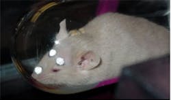
To prepare a mouse for imaging with AwakeSPECT, it is first tagged with three markers that are glued to its head for the infrared system to track. Once the radionuclide is injected, the mouse can then be imaged as it rests in a burrow-like, clear tube. The beauty of the system is that it doesn't require that the mouse remains motionless.
Weisenberger says the next step is to improve the AwakeSPECT imager by upgrading the infrared tracking system, using newer technology for the SPECT imager, and by making the system more intuitive for animal researchers to operate.
Two patents have been awarded to Jefferson Lab for the innovative technology associated with the system.
Related items from Vision Systems Design that you might also find of interest.
1.Miniature microscope helps image mouse brain
A miniature microscope weighing less than 2 grams and tiny enough to balance on the tip of a finger has been developed by Stanford University (Stanford, CA, USA) researchers.
2.UK researchers to shrink MRI system
A new system that enables medical personnel to image difficult areas of the body is being developed by researchers at Imperial College London (London, UK).
3.Algorithms lower the dose of CT scans
Software algorithms developed by US researchers have enabled a new generation of CT scanners to be built that expose patients to one-fourth the radiation of conventional CT scanners.
-- Dave Wilson, Senior Editor,Vision Systems Design