MIT develops hand-held scanner for detecting eye disease
Researchers from the Massachusetts Institute of Technology (MIT) have developed a hand-held ophthalmic-screening instrument that enables the early detection of eye diseases, including diabetic retinopathy, glaucoma and macular degeneration.
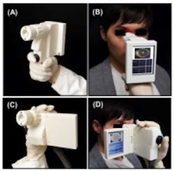
In paper published in Biomedical Optics Express, MIT researchers say that the hand-held scanning device combines 3D imaging, a tiny micro-electro-mechanical systems (MEMS) mirror for scanning, and a technique to correct unintentional movement by the patient. With these technologies, clinicians will be able to collect data with a single measurement, according to MIT, via The Optical Society.
The new device utilizes optical coherence tomography (OCT), which the MIT group helped to develop in the early 1990s. OCT sends beams of infrared light into a patient’s eye and onto the retina, and echoes of this light return to the instrument, which uses interferometry to measure changes in the time delay and magnitude of the return echoes, revealing the cross sectional tissue structure of the retina. This technique is similar to radar or ultrasound imaging, according to The Optical Society.
In what is essentially a shrunken version of a current OCT imager that uses a MEMS mirror to scan the OCT imaging beam, the new device was tested out in two separate designs, both of which were able to acquire images comparable in quality to conventional table-top OCT instruments used by ophthalmologists. To compensate for motion, the instrument takes multiple 3D images of the same area at once, but with different scanning directions. By using these images, the researchers were able to correct distortions and extract images.
The next step for the handheld device is testing in a clinical setting, but researcher and co-author of the paper James Fujimoto says that before the technology finds its way into a physician’s office or in the field, manufacturers would have to find a way to support or lower its hefty cost.
View the research paper.
View the Optical Society press release.
Also check out:
MIT and Massachusetts General Hospital developing new X-ray imaging technology
Camera module and image processor device aids visually impaired
(Slideshow) 10 different ways 3D imaging techniques are being used
Share your vision-related news by contacting James Carroll, Senior Web Editor, Vision Systems Design
To receive news like this in your inbox, click here.
Join our LinkedIn group | Like us on Facebook | Follow us on Twitter | Check us out on Google +
About the Author

James Carroll
Former VSD Editor James Carroll joined the team 2013. Carroll covered machine vision and imaging from numerous angles, including application stories, industry news, market updates, and new products. In addition to writing and editing articles, Carroll managed the Innovators Awards program and webcasts.