Page 2: Handheld microscope could identify cancer cells
Editor's note:This article is continued from page one.
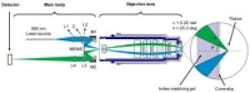
The method used actively aligns the illumination and collection beams in the microscope through the use of a pair of rotatable alignment mirrors. Incorporation of a custom objectivelens, with a small form factor for in vivoclinical use, enables the device to achieve an optical-sectioning thickness and lateral resolution of 2.0 and 1.1 µm, respectively.
For preliminary assessment of the optical design, and to evaluate the performance of the microscope, an ORCA Flash 4.0sCMOS camera from Hamamatsu with a 2D detector was used to mimic the 1D linear detector will be incorporated in the final design. The ORCA Flash features a 16-bit 2560 x 2160 sCMOS image sensor that can achieve a frame rate of 100 fps in Camera Link, or 30 fps in USB 3.0.
A thin rectangular region of interest within the sCMOS array serves as a digital slit, as described previously for a large-scale tabletop LS-DAC microscope system. The camera enabled the rapid acquisition of a 2048 x 8 pixel region at the center of the camera. Since the pixel spacing is 6.5 μm, the team binned the center three rows of pixels to create a digital slit of thickness 6.5 × 3 = 19.5 μm., which approximately matches the diffraction-limited FWHM spot size of ~15 μm that is expected at the detector. Exposure times of 125 μs (a line-acquisition rate of 8 kHz), according to the paper, utilized to collect 500 lines per frame at an imaging rate of 16 fps. In summary, the device images a FOV of 300 x 300 μm with a sampling density of 500 × 600 pixels (x by y).
Researchers expect to begin testing the device as a cancer-screening tool in clinical settings next year, with the hopes that it can be introduced into surgeries or other clinical procedures within the next two to four years.
"For brain tumor surgery, there are often cells left behind that are invisible to the neurosurgeon. This device will really be the first to let you identify these cells during the operation and determine exactly how much further you can reduce this residual," said project collaboratorNader Sanai, professor of neurosurgery at the Barrow Neurological Institute in Phoenix. "That’s not possible to do today."
The research was funded by the National Institutes of Health through its National Institute of Dental and Craniofacial Research and National Cancer Institute.
View theUW press release.
View the academic paper.
Share your vision-related news by contactingJames Carroll, Senior Web Editor, Vision Systems Design
To receive news like this in your inbox,click here.
Join ourLinkedIn group | Like us on Facebook | Follow us on Twitter
Page 1 | Page 2

About the Author

James Carroll
Former VSD Editor James Carroll joined the team 2013. Carroll covered machine vision and imaging from numerous angles, including application stories, industry news, market updates, and new products. In addition to writing and editing articles, Carroll managed the Innovators Awards program and webcasts.