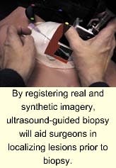
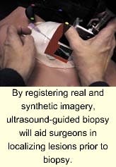
In recent years, ultrasound-guided biopsy of breast lesions has been used for diagnostic purposes, partially replacing open surgical intervention. The procedure also is used for localizing lesions prior to biopsy and cyst aspiration, but for such interventions, however, it is difficult to learn and perform.
In such procedures, good hand-eye coordination and three-dimensional visualization skills are needed to guide the biopsy needle to the target tissue area. Accordingly, University of North Carolina (Chapel Hill, NC) researchers, guided by Prof. Henry Fuchs, are developing an augmented-reality (AR) system that combines computer graphics with video images. The system uses ultrasound echography imaging, laparoscopic range imaging, a video see-through head-mounted display (HMD), and a graphics workstation to create live images that combine computer-generated imagery with the live patient images.
"Such AR systems displaying live ultrasound or laparoscopic data in real time could assist and guide physicians during surgical procedures," says Fuchs. Although HMDs for augmented reality are not commercially available, researchers modified a Glasstron HMD from Sony (New York, NY) that provides SVGA resolution. For optoelectronic tracking of the HMD, two IK-SM43H miniature video cameras from Toshiba Imaging Systems Division (Irvine, CA) were mounted on a wing-like aluminum rig with three IR light-emitting diodes.
Based on a Reality Monster workstation from Silicon Graphics Inc. (Mountain View, CA), imagery from the HMD cameras and the ultrasound imager is transferred into the systems' memory. While the camera video is displayed in the background, ultrasound video images are transferred into texture memory and displayed inside a synthetic opening within the scanned patient. The display presented to the user is updated at 10 Hz and provides ultrasound slice display and rendering of up to 16 million pixels.
