3D imaging technology could improve colonoscopy procedures
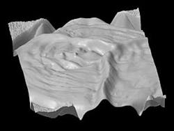
Researchers from MIT have developed an endoscopy technology called photometric stereo endoscopy (PSE) that captures topographical images of the colon surface to create 3D images which provide a more robust way to screen for precancerous lesions of the colon.
Colorectal cancer kills approximately 50,000 people per year in the United States and early detection methods of lesions and polyps have shown to reduce overall death rates. With photometric stereo endoscopy, detection rates could feasibly become even higher. Nicholas Durr, a research fellow at the Madrid-MIT M+Visión Consortium and the Wellman Center for Photomedicine at the Massachusetts General Hospital in Boston, said in an MIT press release that with PSE, precancerous lesions will be more easily detected.
“In conventional colonoscopy screening, you look for these characteristic large polyps that grow into the lumen of the colon, which are relatively easy to see,” Durr said. “However, a lot of studies in the last few years have shown that more subtle, nonpolypoid lesions can also cause cancer.”
Durr, who is the senior author of a paper describing PSE, continued, ““Photometric stereo endoscopy can potentially provide similar contrast to chromoendoscopy,” Durr says. “And because it’s an all-optical technique, it can give the contrast at the push of a button.”
PSE reproduces the topography of a surface by measuring the distances between multiple light sources and the surface. Those distances are used to calculate the slope of the surface relative to the light source, generating a representation of any bumps or other surface features, but this technology had to be modified for endoscopy due to the fact that it is impossible to determine the precise distance between the tip of the endoscope and the surface of the colon. As a result, images generated during the initial attempts contained distortions, particularly in locations where surface height changed gradually.
The researchers filtered out spatial information from the smoothest surfaces, and as a result, the PSE technique does not require the calculation of exact height or depth of surface and instead creates a visual representation that allows for the colonoscopist to determine if there is a lesion or polyp.
Both prototypes developed by the team successfully created 3D representations of polyps and flatter lesions. The researchers plan to test PSE technology in human patients in clinical trials at Mass General Hospital and the Hospital Clinico San Carlos in Madrid. In addition, they are working to develop algorithms that will help to automate the process of polyp and lesion identification.
Also check out:
3D imaging machine helps physicians identify cancer earlier, more frequently
Google Glass making its way into hospitals
How robots could remove blood clots with medical imaging and steerable needles
Share your vision-related news by contacting James Carroll, Senior Web Editor, Vision Systems Design
To receive news like this in your inbox, click here.
Join our LinkedIn group | Like us on Facebook | Follow us on Twitter | Check us out on Google +
About the Author

James Carroll
Former VSD Editor James Carroll joined the team 2013. Carroll covered machine vision and imaging from numerous angles, including application stories, industry news, market updates, and new products. In addition to writing and editing articles, Carroll managed the Innovators Awards program and webcasts.
