Ultrasound imager generates real-time performance
Nondestructive ultrasonic testing of structural components is often used to provide information on subsurface faults in both thick and thin materials. In conventional C-scan ultrasound technology, images are generated by mechanically scanning a probe across the target. After the scan is completed, the test system generates a two-dimensional image.
According to Robert Lasser, vice president of Imperium (Rockville, MD), such methods are often too slow and difficult to use. To overcome these limitations, Imperium has developed a C-scan system capable of generating ultrasound images at 30 frames/s. Typical imagery has a spatial resolution of approximately 0.2 mm.
Company engineers built a 128 x 128-pixel piezoelectric sensor using the same fabrication technology used to develop infrared (IR) sensors. This approach allows the sensor to detect ultrasound images in the 1- to 10-MHz frequency range.
To generate C-scan images, ultrasound is introduced into the target through a large, unfocused transducer that is controlled by a PC. Scattered energy is then collected by an acoustic lens, built by Imperium, and focused onto an ultrasonic array. "The use of a lens provides a simple, inexpensive alternative to complex beam-forming methods often used in ultrasound imaging," says Lasser. During operation, images are focused by adjusting the lens while observing the image. This approach also provides a means to trade off resolution and area coverage.
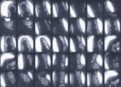
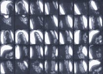
RIGHT. Using Imperium's novel ultrasound imager, ultrasound images, such as those shown of a human finger, were taken at 5 MHz and captured at 30 frames/s.
Each time an ultrasound signal is sent into the material, analog signals from the ultrasound array are read out and digitized using a TMS320C44-DSP, PCI-based frame grabber from Coreco Inc. (St-Laurent, Quebec, Canada). This DSP is programmed to perform nonuniformity correction and brightness/contrast enhancement. After image processing, images are transferred over the host PCI bus to the PC's graphics controller, where images are displayed at 30 frames/s.
In medical imaging, complex beam forming generates images perpendicular to the surface of the target. The real-time C-scan is generated parallel to the target. The Imperium-based imagery method also does not exhibit any speckle typically taken with ultrasound imaging. This means imagery of physical structures can be seen in a higher resolution than that of current ultrasound imaging and without any radiation. Applications of this technique include real-time evaluation of tendons, evaluation of breast cancer, and real-time guidance of biopsies.
Imperium's Acoustocam system is several orders of magnitude faster than currently available systems. "If an inspection is performed of a 10 x 10-ft component with a minimum spatial resolution of 21 mils and a scanning rate of 12 in./s," says Lasser, current C-scan technology would require 6096 passes and take 16.9 h. "Using real-time C-scan technology, only 48 passes would be required, and the task would be accomplished in eight minutes," he adds.
During the development of the technology, Imperium has recorded images of medical structures, faults in metals, composites, and voids inside chips. To view these images, the company has supplied a number of video segments on its Web site: www.imperiuminc.com/.

