Snapshots
Rescuing recorded sound from silence
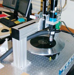
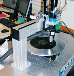
While listening to National Public Radio (Washington, DC; www.npr.org), Carl Haber, a physicist with Lawrence Berkeley National Laboratory (LBNL; Berkeley, CA, USA; www.lbl.gov), learned that librarians at the Library of Congress (Washington, DC, USA; www.loc.gov) were faced with a problem. Much of the library’s audio collection is recorded on wax cylinders and shellac and lacquer disks, many of which are now too fragile to play in their original format.
“I knew sound was stored mechanically and realized images could be used to recreate the sound,” says Haber. Since he already had access to a machine that made high-resolution digital scans, he realized that an optical scanning system could solve the library’s problem.
Haber used a beam of light to illuminate a record’s surface. Flat bottoms of the grooves and the spaces between tracks appeared white, while the sloped sides of the grooves, scratches, and dirt looked black. The image was then computer analyzed. The program found the edges of each groove by focusing on areas of high-contrast and corrected areas where scratches, breaks, or wear made the groove wider or narrower than normal.
On antique monaural disks, sound is recorded in horizontal wiggles of the record groove. On cylinders, sound is recorded in the vertical plane-the depth of the groove. “With disks, we used a camera to image the disks in two dimensions. Once we understood how cylinders were recorded, we needed to measure the third dimension.” Haber says.
With the library’s urging, Haber produced a dedicated disk scanner, IRENE, that was installed at the library last summer. The current 3-D scanning process takes 20 hours to record one minute of sound. But a new version of the confocal scanner, developed for the dental industry, should reduce this time to about 10 minutes. Sound reproduction from the scanner can be heard on-line at http://irene.lbl.gov.
Laser scanning helps blow-mold manufacturing
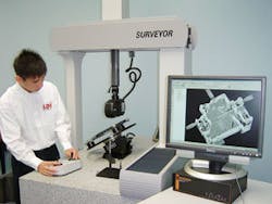
Uniloy Milacron (Tecumseh, MI, USA; www.uniloy.com), a provider of blow-molding systems, recently found that coordinate-measuring machines (CMMs) limited the accuracy achieved in reproducing an existing bottle or model. The company turned to service bureau provider GKS Inspection Services (Plymouth, MI, USA; www.gks.com), a division of Laser Design (Minneapolis, MN, USA; www.laserdesign.com) for assistance.
GKS used the Surveyor DS Series 3D laser scanning system from Laser Design to digitize a 16-oz single-serve dairy container that was too soft to accurately digitize with a CMM. GKS engineers scanned the part, generating a point cloud with the millions of points needed to accurately define the complex surface geometry of the part.
The DS Series laser projects a line of light about 2 in. long onto the part and captures coordinates along this line at scanning rates up to 75,000 points/s. The part is also rotated on the four-axis part-holding fixture that has an integrated rotary stage. This programmable rotation allows for surfaces not viewable from any one angle to be exposed for scanning at a more suitable orientation.
The point cloud is then processed into a NURBS Surface CAD Model using Geomagic STUDIO software. The model is output via IGES or STEP formats into Uniloy’s CAD/CAM system for further modification or manufacturing into a blow mold with no changes. Each point was accurate within 20 µm, and the surfaces generated from the point cloud were accurate to at least 0.004 in. GKS used Raindrop Geomagic software to convert the point cloud to a surface model. It then exported the surfaced model as an IGES file and imported it into CATIA, one of the CAD packages used by Uniloy.
CVPR ’07: Bringing computer vision into focus
At the IEEE Computer Society Conference on Computer Vision and Pattern Recognition (CVPR; cvpr.cv.ri.cmu.edu) this year, Microsoft Research (Redmond, WA, USA; research.microsoft.com) presented papers highlighting research in computer vision. In“Removal of image artifacts due to sensor dust,” Changyin Zhou of Shanghai Fudan University (Shanghai, China; www.fudan.edu.cn) and Steve Lin, a researcher for Microsoft Research Asia, offered a technique to remove dust effects from digital photographs automatically. “Currently,” Lin says, “dust effects in photographs must be removed by hand using software. We provide an automatic tool for dealing with this problem. The key idea is to model the physical formation of dust artifacts and obtain results superior to those of general artifact-removal techniques.”
In “Inferring temporal order of images from 3D structure,” Sing Bing Kang, a senior researcher at Microsoft Research, discussed a technique to order chronologically a large set of photographs by analyzing the presence of structures visible in the photographs. “Given a large collection of historical photographs of a city that spans decades or perhaps even a century,” Kang says, “how can photographs automatically be sorted by time? We attacked the problem by reasoning about structures visible in photographs. This is made possible by assuming that each structure is unique and exists in a continuous block of time. Sorting photographs in time,” Kang says, “allows time-varying 3-D models to be constructed from collections of historical photographs. The user can then navigate in space and time. ”
Faces reveal genetics
“Many genetic conditions affect the development of the face,” says professor Peter Hammond of the UCL Institute of Child Health (London, UK; www.ich.ucl.ac.uk). “While faces characteristic of Down’s syndrome can be easily recognized, there are other, rarer conditions such as Williams syndrome 7. This makes the temples narrower, the mouth fuller, and the jaw smaller.”
Hammond has developed software that compares a 3-D picture of a child’s face with other faces, to see which abnormal face fits most closely. The image of the abnormal face is a composite of between 30 and 150 children with that condition. Each image contains approximately 25,000 points on a face surface, capturing contours in 3-D.
Hammond envisages a future in which doctors take 2-D pictures of children’s faces and e-mail them to a specialist center that converts them to 3-D, compares them with model faces, and e-mails back suggestions about which genetic tests to perform.


