An easy camera to swallow
Diseases of the small bowel or small intestine are notoriously difficult to diagnose, have vague symptoms, and are usually advanced at the time of definitive diagnosis. Current methods, including endoscopy and radiological imaging, permit visualization only of approximately the first one-third of the small bowel. Such procedures can be expensive, produce limited results, and cause discomfort for patients. What is needed is a noninvasive, easy-to-use diagnostic tool that provides an improved level of visual imaging.
Photobit (Passadena, CA) has designed a tiny camera that can be swallowed like a pill and can take images of the stomach and small bowel as it passes through them unaided. As an ingestible capsule, the 3-mW sensor, which measures 1 x 30 mm, produces color video of the gastrointestinal (GI) tract.
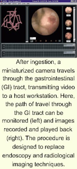
Dubbed the M2A disposable imaging capsule and marketed by Given Imaging (Atlanta, GA), the device incorporates a miniature light source, transmitters, and power cells. After the capsule is swallowed by the patient, it moves with the aid of peristalsis through the GI tract. As the capsule passes through, it transmits video signals that are stored in a receiving unit, which enables the system to trace the physical course of the capsule's progress. A wireless recorder worn on a belt around the patient's waist receives signals transmitted by the capsule through an array of antennae placed on the patient's body. The ambulatory belt permits patients to continue about their daily activities during the GI examination until the capsule is excreted naturally from the body.
A computer workstation, equipped with reporting and processing software processes the data and produces a short video clip of the small intestine together with additional data from the digestive tract. The workstation permits physicians to view, edit, and archive the video and save individual images and short video clips.
"Our goal has always been to commercialize a minimally invasive, disposable imaging capsule for diagnosing small-intestine disorders," said Dr. Gavriel Meron, president and CEO of Given. "Photobit's sensor, which combines all camera functions on a single chip, has made this a reality." At present, the device is not for sale in the United States and is limited by federal law to investigational use.

