Workstation software speeds craniofacial analysis and visualization
Using Pentium-III, Windows-NT-based workstations and graphics accelerators, optimized visualization software results in detailed 3-D analysis of anatomical structures.
By R. Winn Hardin,Contributing Editor
During the past few years in the medical field, computer workstations supplied by SGI (Mountain View, CA) have been the preferred platforms for structuring intensive three-dimensional (3-D) imaging systems, such as craniofacial protocols, using computed-tomography (CT) and magnetic-resonance-imaging (MRI) technologies. The workstation multimedia design uses custom connections for increased bandwidth between the microprocessor and the graphics card. This approach has proved effective for technical and medical imaging systems where a single imaging session can generate up to 1000 512 x 512 x 16-bit images. Optimized hardware for visualization workstations comes with a high price, however.
FIGURE 1. Physicians, radiologists, and imaging specialists at universities throughout North America, including the Universities of Iowa and Minnesota, have helped Vital Images evaluate and develop its Vitrea 2 three-dimensional visualization software for craniofacial and other medical imaging protocols. An operator at the University of Iowa reviews a 3-D volume image of a skull.
To compete cost-effectively with available visualization systems, engineers at Vital Images (Minneapolis, MN) have designed Vitrea 2 medical visualization software. They have combined optimized code, dual-processor Intel Corp. (Santa Clara, CA) Pentium-III, Microsoft (Redmond, WA) Windows-NT-based workstations, and graphics accelerators from 3Dlabs (Sunnyvale, CA) to derive improved performance for 3-D visualization applications.
Vital Images has qualified its
Vitrea 2 software on both the Hewlett-Packard (Palo Alto, CA) Kayak and the SGI 320 visualization workstations—both dual-Pentium III, 500- to 600-MHz Windows-NT platforms (see Fig. 1). "There are a lot of equivalents out there that we'll be qualifying later," explains Vital Images founder and chief technical officer Vincent Argiro. Vital Images also plans to support Vitrea 2 on the SGI O2 platform. However, Argiro maintains that the performance of the HP Kayak Windows-NT system with dual Pentium IIIs and a GVX1 accelerator card from 3Dlabs outpaces the SGI O2 workstation performance, meets or beats the performance of the multimedia SGI 320 dual-processor visualization workstation, and costs less than both.
Streaming extensions
The new Windows NT systems, Argiro says, offer parallel processing and streaming single-instruction multiple-data extensions (SSEs). These extensions allow the same instruction set to be performed quickly on large data sets. Integrating Vitrea 2 software into these systems means that billions of pixels contained in large CT or MRI applications, which often contain as many as 1000 image slices of the patient, can be quickly and efficiently rendered into 3-D voxels for diagnosis or preoperative planning.
FIGURE 2. Several computer workstation platforms, including the SGI O2, SGI 320, and Hewlett-Packard (HP) Kayak, can run Vitrea 2 visualization software. Vital Images engineers claim that the HP Kayak Windows-NT-based workstation provides the fastest performance, thanks to the Intel Pentium III's special handling of long data streams.
Explains Argiro, "Intel has realized what SGI really pioneered five years ago, which is an understanding that in many visualization applications, data movement is just as, or more, important than data manipulation or arithmetic. It takes careful consideration of how the data are moved, in what sequence, and how to take advantage of opportunities where you can handle data in parallel. In most applications [with visualization], you're dealing with groups of short vectors, such as RGB or x-y-z. So the SSEs were an ideal vehicle for this application, just as similar methods have been applied to many graphics applications by others in the industry."
FIGURE 3. Vitrea 2 software report screen allows a technician to create reports using images saved in the Viewer window. These reports can be printed to a Postscript printer or a laser printer, or can be posted to a secured intranet through a network interface card. These multiple views, each with different anatomical features, are of the same lesion located just below the patient's jaw.
In addition to optimizing algorithms through the SSE instruction set, Argiro and his associates have worked closely with 3Dlabs on its new Oxygen GVX1 accelerator card (see Fig. 2). "We've done a lot of work with 3Dlabs to optimize the driver of that card for this kind of system. The cards are targeted at the technical imaging market, not just the gaming side, even though the two markets are coming together," Argiro claims.
The GVX1 is a less-than $1000 accelerator card designed specifically for workstation use that is fully OpenGL compatible and capable of handling 246 Mbytes of textures in on-board hardware with 30 times that amount of virtual texture memory space. 3Dlabs offers PowerThread functionality to maximize the Pentium-III SSE instruction set, balancing lighting and texture operations between the Gamma on-board processor and the two Pentium IIIs on the Hewlett-Packard Kayak motherboard. The board comes with a 15-pin analog monitor port or a digital DFP (digital flat panel) port and is compatible with composite or S-Video, NTSC, and PAL formats.
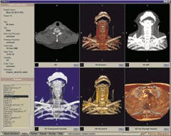
FIGURE 4. Various image-processing and display functions are called into play based on physician/patient needs. This gallery of separate views demonstrates various surface rendering, shading, and sizing of images. Other tools are automatically generated through the selection of the Carotid CT Protocol.
At present, dual monitor systems are only available with the SGI O2 Unix-based system. Argiro says that software allowing dual-monitor operation for the Windows NT environment is expected later this year. Vitrea 2 software is available on the SGI 21-in., 1280 x 1024-pixel, double-buffered, 32-bit monitor. Another advantage of the Windows-NT environment is its ability to save the final 3-D-rendered images as a JPEG file for inclusion in PowerPoint files or other productivity tools in addition to the DICOM 3.0 file standard supported by Vitrea 2 for communications among picture-archive-and-communication (PAC) systems and medical workstations (see Fig. 3).
Vital Images recommends 1 Gbyte of RAM for both the SGI 320 and HP Kayak workstations. According to Argiro, competing systems use 1 Gbyte or more of memory but fail to render 3-D models from 1000-slice CT or MRI applications in a single pass through the platform. He says, "Often, the optimization of the algorithms hinges on a few key observations of the most commonly used [software function]. We were down to counting the number of cycles that were used on the Pentium to render each voxel before handing it off to the graphics hardware. We've looked closely on our own and with SGI and 3Dlabs to provide a level of tuning that most organizations frankly don't have the capability to do" (see Fig. 4).
Both the SGI and HP workstations also accommodate two 18-Gbyte internal hard drives. The HP workstation adds an Adaptec (Milpitas, CA) SCSI RAID interface/controller card for fast loading and redundant storage of data. The Vitrea 2 workstations also come with a 10/100Base-T Ethernet network interface card and an optional CD-ROM drive for data backup. Vitrea 2 can save files in DICOM (digital imaging and communications in medicine) 3.0 or as an HTML (hypertext markup language) document for posting to a secured hospital intranet.
Image-processing tools
Says Argiro, "The Circle-of-Willis protocol was among the first and most popular automated protocols we offered. Most users don't have to adjust any settings. The volume rendering sets a shiny surface [shade function] to look at the bone and blood vessels, a color coating [contrast function] from white for blood vessels in dense areas of the skull, and orange and red for vessels where the contrast is less dense. It can also automatically narrow down the field of view along the skull [trim function]. It can call up quantitative tools such as rules to allow measurement of the diameter or volume of an aneurysm. It also makes it easy to document or publish those works. These features are used most often by neurosurgeons, even though they only represent about 10% to 15 % of the functionality of the product" (see Fig. 5).
In addition to area, volume, distance, and angular measurements, Vitrea 2 also offers maximum/minimum intensity projection rendering, several fly-through options, and click-and-point snapshots for easy report generation. Other functions include several kinds of multiplanar reformatted views (three orthogonal planes converging on one spot) with 3-D control of the three planes to allow cross sections of diagonal or curved internal structures; digital movie creation; batch operations; digital annotation with arrows and cross hairs; and easy correlation between 2-D CT/MRI slices and three-dimensional renderings.
Company Information
Adaptec Inc.
Milpitas, CA 95035
(408) 945-8600
Fax: (408) 262-2533
Web: www.adaptec.com
Hewlett-Packard Co.
Palo Alto, CA 94304
(650) 857-1501
Fax: (650) 857-5518
Web: www.hp.com
Intel Corp.
Santa Clara, CA 95052
(800) 548-4725
Web: www.intel.com
SGI
Mountain View, CA 94043
(650) 960-1980
Web: www.sgi.com
3Dlabs Inc.
Sunnyvale, CA 94086
(408) 530-4700
Fax: (408) 530-4701
Web: www.3dlabs.com
Vital Images Inc.
Minneapolis, MN 55416
(612) 915-8009
Fax: (612) 915-8010
Web: www.vitalimages.com