Researchers monitor nanofiber spinning with CCD camera and zoom optics

July 16, 2010
The University of British Columbia's Advanced Fibrous Materials Laboratory is developing polymer-based nanofibers with the assistance of a CCD camera equipped with a Navitar lens for monitoring fiber properties during the electrospinning process.

Led by fiber materials expert Frank Ko, PhD, the Advanced Fibrous Materials Laboratory (AFML), a research laboratory at the University of British Columbia (UBC; Vancouver, BC, Canada) in the department of Materials Engineering, conducts research in the areas of structural, functional, and biomedical electrospun fibers.

With its nanofiber R&D focus on the medical field, the AFM team is working to enable advances in artificial organ components, surgical implants, vascular stents, drug delivery, wound dressings, medical textile materials, and tissue engineering scaffolds in orthopedic, vascular, and neural prostheses. The nanofiber in development is based on polymer solutions that use organic and inorganic nanoparticles. Properties such as the high surface-to-volume ratio and a relatively defect-free structure on the molecular level are what support the mechanical strength and chemical suitability of nanofibers in these composite material applications.1
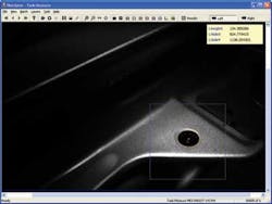
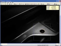
Inside a nanofiber electrospinning unit, a CCD camera equipped with a Navitar Zoom 6000 lens helps the researchers monitor the polymer solution and fine-tube the nanofiber properties. (Photo courtesy of AFML)AFM employs Navitar (Rochester, NY, USA) optics during an electrospinning process, which uses an electric potential between 5 and 10 kV to draw very fine fibers from a liquid (a polymer solution). Precise control of the fiber creation process is critical to enabling the properties that allow the nanofibers to be used in medical applications. During the initial stages electrospinning, flow rate and desired potential must be balanced. To vary the flow rate of the polymer solution, AFM must observe the jet emerging from the nozzle and the tip of the syringe that contains polymer solution, which can be difficult. A CCD camera equipped with a Navitar Zoom 6000 lens is used to monitor the polymer solution and fine-tune the fiber properties. References1. http://en.wikipedia.org/wiki/Electrospinning-- Posted by Vision Systems Design

Voice Your Opinion

To join the conversation, and become an exclusive member of Vision Systems Design, create an account today!