EEG monitor uses bi-spectral analysis to optimize anesthesia
EEG monitor uses bi-spectral analysis to optimize anesthesia
By Lawrence H. Brown, Contributing Editor
Ten years ago, while he was studying biomedical engineering at Boston University (BU; Boston, MA), Nassib Chamoun became interested in applying bi-spectral analysis to medical functions. Chamoun applied the method to electroencephalogram (EEG) data and left BU to found Aspect Medical Systems (Natick, MA).
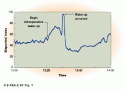
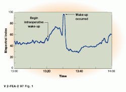
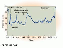
Aspect?s A1050, which has received FDA approval, interprets EEG data using bi-spectral analysis and displays a bi-spectral index (BIS index) that allows doctors to study the results of anesthesia in real time. Ranging from 0 to 100, an index between 60 and 100 represents consciousness, while values below 60 indicate various states of unconsciousness (see Fig. 1). Using the BIS index anesthesiologists get information that allows them to administer lighter doses of medication, enhancing the safety of patients and the efficiency of operations. Using the index, anesthetic dosage can be 25% less than is normally given, saving thousands of dollars in postoperative costs because wakeup times are faster. And, with a real-time view of the patient?s state of consciousness, dosage reactions and equipment irregularities can be seen clearly and quickly (see Fig. 2).
Most EEG analyzers use power spectral analysis to measure EEG signal amplitude as a function of frequency. But because power spectral analysis cannot measure relationships that exist among brain waves, only a linear interpretation of the waveforms from each side of the brain is possible. Bi-spectral analysis tracks changes in signals arising from linear and nonlinear changes in waveforms, and subtle changes in brain activity and medication dosage become highly quantifiable (see OBi-spectral analysis makes medication more manageable,O p. 18).
When designing the A1050, the Aspect team recognized the need for accurate digitization and real-time computation. To provide these features, a combination of custom signal converters and off-the shelf processors were married with a real-time operating system. In this way, Fourier and bi-spectral analysis of EEG waveforms could be computed and the resulting BIS index displayed in real time.
Reducing noise and distortion
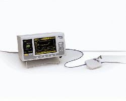
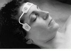
To obtain a high signal-to noise ratio, EEG electrodes were designed as a self-adhering band and slotted for placement of pregelled, disposable electrodes. This reduced source impedance and helped match the impedances from the electrodes to the system. Attached to the electrodes is a signal converter that is clipped to a bedsheet or placed under a mattress. Digitizing the signal as close to the patient as possible minimizes extraneous interference from the patient to the converter and isolates the converter from the noise of the monitor?s digital signal processor (see Fig. 3).
Low noise and distortion were mandatory in rendering signals capable of computational analysis. OThe problems encountered in filtering noise and distortion are similar to those in the audio industry,O says John Shambroom, director of engineering. Using audio-industry models, he designed a sigma-delta network converter for EEG data acquisition. In operation, analog signals are digitized by this converter and multiplexed into a serial data stream, which outputs 1-bit samples at 16,384 samples/s.
The signal converter is attached to the system?s processor by a 20-ft
cable. This unit houses the CPU motherboard, video screen, and a
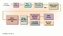
display that shows the BIS index. Three processors are used in the A1050: an Intel 80286, a Texas Instruments TMS 320C30, and an Analog Devices ADSP 2105. In operation, the ADSP 2105 filters, reconstructs, and reduces EEG data. These data are then output to the TMS 320C30 that computes the BIS. The Intel 80286 controls the video display, keyboard, and serial port (see Fig. 4).
The A1050 is also programmed to reduce spurious signals from the
patient or other sources in the operating room that distort EEG data. These artifacts, which occur when patients blink or move, are filtered using software running on the host CPU. Once frequencies are filtered and adjusted, EEG data are transformed into the frequency domain. OBi-spectral algorithms then determine the phase relationships of the individual EEG signals,O says Rick Noonan, director of product development.
Multitasking in real time
For the operating system (OS), VRTX, an off-the-shelf, real-time, multitasking system from Microtec Research (Santa Clara, CA), was used. OVRTX was chosen for its range of services that include mailboxes for passing messages between tasks, semaphores that control access to restricted resources, and event flags that let tasks know an event has happened,O says Noonan.
VRTX OS applications are composed of several tasks that are small, self-contained programs. These tasks execute at a frequency based on their priorities and the events driving them. Communication between tasks is handled by the OS services. Coding applications as tasks under a real-time multitasking OS, rather than writing one program that performs every task, simplified software development. ODesigners need only choose optimum task priorities and communication mechanisms to solve problems such as processor loading, process synchronization, and real-time response,O says Noonan. VRTX?s context-switch time and interrupt latency and the system?s maturity contributed to the decision to use VRTX.
On-board boot code for the three processors includes code that tests the system?s hardware and loads the main programs. Because of continued feature enhancements, updated algorithms are programmed in EEPROMs and mailed to customers every six months.
EEG and EKG
Bi-spectral analysis techniques Bi-spectral analysis techniques can be applied to many different areas of signal and image processing to extract anomalies in data that may not otherwise be seen. Because of the success of the technique in EEG analysis, Aspect plans to use bi-spectral analysis in the design of an electrocardiogram (EKG) analyzer. Currently under development, the machine will allow physicians to more closely monitor heartbeat patterns.
FIGURE 1. BIS index is elevated when a patient is intentionally roused to test motor function during spinal surgery. The peak in the BIS is followed by a return to a deeper sleep state.
FIGURE 2. An unexpected rise in the BIS index alerts the anaesthesiologist to a failure in a pump battery used to deliver anesthesia. Toward the end of the surgery, the BIS index shows the effects of the anesthesia wearing off and the patient`s return to consciousness.
FIGURE 3. To increase the signal-to-noise ratio of the EEG analyzer, signals are digitized as close to the patient as possible using a portable signal converter. This is attached via a 20-ft cable to the EEG analyzer.
FIGURE 4. EEG analog signals are digitized in a sigma-delta converter and then multiplexed to a serial 1-bit, 16,384-samples/s data stream. In the system, a Texas Instruments TMS320C30 filters, reconstructs, and reduces the data before computation of the bi-spectral index by the host CPU.
Company Information
Analog Devices
One Technology Way
Norwood, MA 02062
(617) 461-3881
Aspect Medical Systems
2 Vision Way
Natick, MA 01760
(408) 980-1300
Microtec Research
Santa Clara, CA
(408) 980-1300
Texas Instruments
Dallas, TX
(800) 477-8924
