Standards steer medical systems toward interoperability
Standards steer medical systems toward interoperability
By Lawrence H. Brown, Contributing Editor
Working as digital, networked systems that store and retrieve images for immediate and simultaneous use, picture archiving and communications systems (PACSs) are now in widespread applications in hospitals and health-care clinics. In the past, the networking of different medical imaging systems proved difficult because of the proprietary image-capture, transmission, and display methods developed by individual manufacturers.
To overcome these problems, a joint committee formed by the American College of Radiology and the National Electrical Manufacturers Association has developed a standard framework known as Digital Imaging and Communications in Medicine (DICOM; see "DICOM sets a medical imaging standard," p. 36). The key to implementing the standard is to build DICOM definitions into imaging systems that bridge hardware devices and software operating systems.
Object-oriented software
One such DICOM application comes from Compurad (Tucson, AZ). Company founder and chief executive officer Philip Berman wanted an easy-to-use lightbox-equivalent digital processing system to view, store, and retrieve radiographic images. To that end, the company developed a PC-based system running under Windows NT/`95. Dubbed Iviewpro, the system uses DICOM 3.0 protocols and mimics a film lightbox by using the entire screen to view the image while eliminating keystroking and minimizing mouse movements.
To develop the software for Iviewpro, Compurad chose Visual C++ from Microsoft (Redmond, WA). By using Active X, a high-level object-oriented interface for Dynamic Linked Libraries (DLLs), components for the Iviewpro program such as viewing images could be built as DLLs. Active X allows these imaging components to be linked together graphically, thereby forming the toolbar-driven menu interface of IviewPro.
"Designing component-based software is like putting together building blocks in construction," says Henky Wibowo, company vice president of engineering. "The components act together to make the complete application successful." Iviewpro`s display feature allows images to be sorted into different stacks to show prior patient studies. "This is useful in comparing studies of multiple images that use different modalities," adds Wibowo.
Improving integration
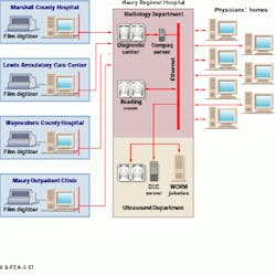
Maury Regional Hospital (Columbia, TN) and Delta Imaging Systems (Nashville, TN) are using Iviewpro to integrate the hospital`s miniPACS ultrasound system and its radiology department. In this PACS, images are transmitted from four remote locations that include two smaller hospitals, two outpatient clinics, and seven physicians` homes (see figure below).
A Proliant 1500 server from Compaq Computer (Houston, TX) acts as the system hub, integrating the ultrasound miniPACS and teleradiology systems. Because this server can separate the data from individual applications, the same data can be accessed by different applications running on different workstations, resulting in improved system cost-effectiveness.
Running under Windows NT/4.0 software, the server uses a synchronized multiprocessor system with two Pentium Pro CPUs, a PCI-bus configuration, and 256 Mbytes of RAM. Inetpro, object-oriented software from Compurad, accesses the data and allows the server to receive and route all the images through a 100-Mbit/s Ethernet line to the network. Windows NT is used to control a RAID system that contains four 4-Gbyte drives, each partitioned into 600 Mbytes. Mirroring these data onto a second drive provides protection from losing data and offers increased data security.
Viewing images
To view images, the hospital`s radiology department uses a reading room equipped with two monitors of 1k x 1k resolution and a diagnostic room containing two monitors of 2k ¥ 2k resolution. Supplied by Image Systems (Minnetonka, MN), the monitors are driven by PCI-bus-based display controllers from Dome Imaging Systems (Waltham, MA) and hosted by Gateway 2000 (North Sioux City, SD) workstations running Windows `95.
Film is digitized by a scanner from Lumisys (Sunnyvale, CA) and is automatically sent to the diagnostic area using Inetpro. Other images from the ultrasound department, such as computed tomography, magnetic resonance, and sonograms, are viewed in the reading room.
Originally, an integrated-services-digital-network (ISDN) line was used to connect the doctors` homes to the hospital, but it proved slow. The ISDN line has been replaced by a fiberoptic cable-modem connection provided by Columbia Cablevision (Columbia, TN). "Using ISDN took 40 minutes to receive a full-chest or pelvic CT scan," says Dr. Richard Stults, radiologist at Maury Hospital. "Now, using a cable-modem line and Iviewpro, that time has been cut to less than three minutes."
Ultrasound integration
The ultrasound miniPACS is the only network that is performing permanent filmless archiving at the Maury Regional Hospital. Four in-house ultrasound systems and Maury`s outpatient clinic send images to a Digital Equipment Corp. (DEC; Maynard, MA) Celebris 6150 network server where the images are processed for storage and retrieval. The DEC server uses a150-MHz Pentium processor, a PCI-bus configuration, and 160 Mbytes of RAM and runs the Unix operating system with NextStep graphical user interface software. ©
Using two monitors in the ultra sound department`s central viewing area, doctors can study images within five seconds after a sonogram is recorded. ALI Technologies (Vancouver, BC, Canada), the systems integrator for the ultrasound miniPACS, provided proprietary software to set up a DICOM association with the Iviewpro application. The software permits accessibility for viewing any image at any time in the hospital, physician`s home, or remote site by routing the images through the Ethernet line to the server.
At the end of the day, the server automatically routes all the images to a Hewlett-Packard (Palo Alto, CA) 80fx optical WORM jukebox. Here, data are written to magnetic optical disks, archiving the data for storage and retrieval. Thirty-two disks provide 2.6 Gbytes of memory. "It`s a secure medium that doesn`t allow the disks to be written over," explains Dana Dryver, a systems sales manager for ALI.
Acceptance of the DICOM standard by vendors is making fully integrated PACSs more realistic. Additionally, the use of the Internet and Java software means that remote users can access data and programs faster and easier, and that the technology becomes portable across different operating systems. The long-term goals of Maury Hospital include transmitting images and patient data electronically to and from intensive care areas, emergency rooms, and different hospitals` laboratories.
Two county hospitals, a care center, and an outpatient clinic, all remotely connected, are hard-wired to the Maury Regional Hospital by a T-1 (1.544-Mbit/s) connection installed by Bell South (Nashville, TN). The Ethernet line transmits images at 10 Mbit/s. Using the Maury Regional Hospital`s picture-archiving and communications system, physicians can transmit and receive x-ray, ultrasound, computed tomography, and magnetic-resonance images across the network.
DICOM sets a medical imaging standard
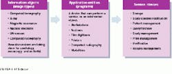
The Digital Imaging and Communications in Medicine (DICOM) standard is intended to promote an open architecture for imaging systems, allowing their interoperability for the transfer of medical images and associated information. Developed by a joint committee formed by the American College of Radiology and the National Electrical Manufacturers Association, DICOM defines information objects (image types such as x-ray or magnetic resonance) and their correspondence to specific images, studies, and reports. The definitions are used to structure a framework in which users and providers of digital imaging equipment, including imaging modalities, picture archiving and communications systems (PACs), and devices such as printers and workstations, can communicate with each other over a networked system, rather than only through point-to-point connections (see figure).
The DICOM standard is organized into two major classes: a service class user (SCU) and a service class provider (SCP). Service class users are devices, such as magnetic resonance imagers, which use the services of other devices, such as workstations and laser printers--service class providers--to display images on a monitor or hard-copy device. Some devices, such as a workstation, can be both a service provider, displaying images, and a service user, sending a command to a printer.
The two major classes are subdivided into service object pairs (SOPs), which are the combination of an information object and a service. In DICOM language, these pairs are essentially specific definitions of how devices request and provide services on a device and form the basis for DICOM conformance.
Eight specific service classes define specific areas to which users or providers must conform--verification, storage, query/retrieve (at four levels--patient, study, series, and image), basic print, advanced print, secure archives, hospital, and radiology information systems (HIS/RIS), and at three distinct levels-- patient, study and results management, and media format.
Companies that want to communicate via the DICOM standard first write a DICOM conformance statement. The conformance statement indicates which optional components are being supported (there are mandatory fields in which all vendors must conform) and which additional components or specializations are being provided.
Conformance statements contain an application data-flow diagram that shows how activities are performed locally and remotely; a detailed specification identifying how the supported SOP classes initiate or accept communications with other devices (also know as an association); options supported under each SOP class (compression with storage service class, for example); communications protocols supported; and other details that relate to DICOM conformance or interoperability.
A major weakness of the DICOM standard is that a vendor needs only to draft a conformance statement to a single service class and a SOP class (verification SCP for computed tomography, for example) and to claim they are DICOM compliant, even though the balance of the SOP and classes may remain undefined. This shortcoming has caused industrywide confusion for vendor conformance regarding the DICOM standard.
Another industrywide dichotomy relates to the costs to implement DICOM-based versus non-DICOM-based solutions. In practice, the DICOM-based solutions are priced higher than their non-DICOM-based counterparts, even if the modality to which the devices are connected provide native DICOM outputs (outputs that do not require the use of a third-party interface device to convert data in a proprietary format to a DICOM file format).
Additionally, less than 20% of the modalities currently in use provide native DICOM output; they therefore require use of a third-party device that can add as much as a $25,000 per modality cost connection to the total system price. This high price tag has had a slight impact in the acceptance and use of the standard, although mainly in the lower end of the PACS` spectrum (teleradiology). Vendors claim the price disparity (native or non-native) is due to their need to recover nonrecurring engineering charges (NREs) related to reviewing and testing their DICOM conformance statement against other companies` conformance statements. However, some vendors disagree with this contention.
Despite its problems, the DICOM standard is an attractive solution to vendors and users alike by allowing disparate modalities to talk a similar language, minimizing the Tower of Babel syndrome that currently exists among modalities. This capability will ultimately expand outward to physicians who will be communicating with these devices through the use of picture archiving and communications systems.
Michael J. Cannavo
Image Management Consultants
A Division of Healthcare Imaging Specialists
Orlando, FL
E-mail: [email protected]
In the Digital Imaging and Communications in Medicine (DICOM) standard, information objects (image types) are recognized by application entities or programs to perform a service class or operation. For example, a magnetic-resonance image (MRI) or a series of MRIs of a patient can be archived for display at a monitor (query/retrieval service). (Diagram courtesy of Kodak Digital Science)


