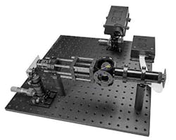
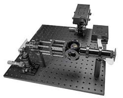
MICROSCOPY: Confocal microscope employs digital light projector
In fluorescence microscopy, specimens are illuminated by a specific wavelength of light that is absorbed by the fluorophores in the sample and emitted at a longer wavelength. In conventional microscopes, this emitted fluorescence can be from a relatively wide focused background area of the sample, with the result that the fluorescence that is required to be measured at a single point is obscured by fluorescence from out of focus points in the sample.
To overcome this interference, confocal imaging techniques increase the contrast of fluorescence images by employing point illumination and a spatial pinhole to eliminate any out of focus fluorescence.
Despite the advantages obtained, such confocal microscopes are expensive since to obtain a 2D image of the sample, it is necessary to scan multiple points across the sample using relatively complex and expensive scanning optics.
To reduce the cost of such systems Aeon Imaging (Bloomington, IN, USA; www.aeonimaging.com) has patented a novel method to perform confocal imaging that uses off-the-shelf optical, opto-electronic and electronic components.
Rather than illuminate the sample with a point light source, the company's Digital Light Microscope (DLM) system employs a LightCrafter digital light projector from Texas Instruments (Dallas, TX, USA; www.ti.com) that is configured to project a series of narrow adjacent lines across the field of view of the sample. This light is then reflected off of a dichroic mirror from Edmund Optics (Barrington, NJ, USA; www.edmundoptics.com) and focused onto the sample using a 20x infinity corrected objective lens from Olympus (Center Valley, PA, USA; www.www.olympusamerica.com).
Returned light from the sample then passes through the dichroic mirror and is focused onto a CMOS-based camera using a tube lens from Mitutoyo America (Aurora, IL, USA; www.mitutoyo.com). Employing a 5Mpixel MT9P031 CMOS sensor from Aptina Imaging (San Jose, CA, USA; www.aptina.com), the camera is operated in rolling shutter mode and synchronized with the illumination source from the LightCrafter digital light projector. Since the light is synchronized with the line light projected from the DLP, light incident on the imager outside the rolling area shutter's exposure area is not exposed.
Just as point light sources in conventional confocal imaging systems increase the contrast of image data captured, so too does this 2D scanning technique. Further, unlike conventional confocal microscopes that require de-scanning of the returned light, this light is directly imaged onto the rolling shutter of the system's CMOS camera, substantially reducing the cost of the system.
According to Dr. Brian McCall, an Optical Engineer with Edmund Optics who designed the 20x fluorescence optical system for Aeon Imaging, the total cost of the components used in the system is approximately $7,500.
Vision Systems Articles Archives

