Imaging technologies married at Purdue
Researchers at Purdue University (West Lafayette, IN, USA) have married two biological imaging technologies, creating a new way to learn how cells metastasize or proliferate, forming a cancerous tumor.
An atomic force microscope (AFM) uses a tiny vibrating probe called a cantilever to yield information about materials and surfaces on the scale of nanometers. The instrument enables scientists to study molecules, cell membranes and other biological structures.
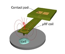
However, the AFM does not provide information about the biological and chemical properties of cells. So the researchers fabricated a metal microcoil on the AFM cantilever. When an electrical current is passed though the coil, it exchanges electromagnetic radiation with protons in molecules within the cell, inducing another current in the coil, which is detected.
Such an advance makes it possible to simultaneously study the mechanical and biochemical behavior of cells, which could provide new insights into disease processes, according to biomedical engineering postdoctoral fellow Charilaos Mousoulis.
A prototype of the system was described in a research paper entitled "Atomic force microscopy-coupled microcoils for cellular-scale nuclear magnetic resonance spectroscopy" that appeared online April 11 in Applied Physics Letters.
The paper was co-authored by Mousoulis, research scientist Teimour Maleki, Babak Ziaie, a professor of electrical and computer engineering and Corey Neu, an assistant professor in Purdue University's Weldon School of Biomedical Engineering.
Related news items from Vision Systems Design.
1. Cell analyzer spots cancer in the blood
An automated optoelectronic image capture and processing system acquires images of cells in the blood and screens them for cancer in real time, with a record-low false positive rate.
2. Oral cancer detected by handheld probe
Researchers led by Dr. John Zhang from the University of Texas at Austin (Austin, TX, USA) have created a portable, miniature microscope that may help reduce the time taken to diagnose oral cancer.
3. Software helps classify skin cancer
Researchers at the University of Edinburgh (Edinburgh, UK) have developed diagnostic software that can help health care workers to correctly identify different types of skin lesions, leading to more effective diagnosis of skin cancer.
-- Dave Wilson, Senior Editor, Vision Systems Design
