3-D scanner captures images of the ear
Measuring the ear canal accurately by taking ear mold impressions is often time-consuming, inefficient, messy and uncomfortable.
Now, based on a principle known as emission re-absorption laser induced fluorescence (ERLIF) that originated in the laboratories of Massachusetts Institute of Technology (MIT, Cambridge, MA, USA; www.mit.edu), engineers at Lantos Technologies (Cambridge, MA, USA; www.lantostechnologies.com) have created a system that can capture ear canal shape.
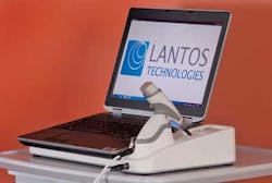
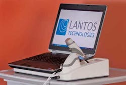
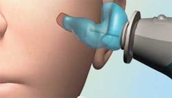
The Lantos 3-D ear scanning system consists of a portable 3-D scanner with a video otoscope tip covered by a soft conforming membrane. In use, the membrane is inserted into the ear guided by a video feed from the otoscope. Once positioned, the membrane is filled with a water-based optical fluorescent dye that expands the membrane until it conforms to the unique ear geometry of the patient.
The otoscope tip is then retracted, and as it is, the fluorescent dye is illuminated by a light source. A fiber-optic camera in the head of the probe then measures the intensity of the reflected light.
After making hundreds of 2-D measurements of the inside of the conforming membrane as the probe tip is retracted, the 2-D geometric data sets are transformed into 3-D data using a patented algorithm. Then they are stitched together in real time to produce a personalized model of the ear canal.
The scanner can also measure the canal wall elasticity by varying the pressure that the membrane exerts on the ear canal wall. This enables important dynamic information about the ear canal wall to be captured in real time.
When the scan is complete - typically in less than a minute - the liquid is drawn out of the conforming membrane and the membrane is completely removed from the ear.
Vision Systems Articles Archives


