Stereo system delivers flicker-free images
Andrew Wilson, Editor, [email protected]
To capture and display three-dimensional (3-D) images, many stereo imaging systems use two cameras to image a scene from two different perspectives. After images are captured they can be displayed using specialized software in either a field or frame sequential manner on autostereoscopic displays. Field sequential broadcast standards such as NTSC dictate the use of a 60-Hz interlaced scan, which means that only 30 stereo pairs/s can be displayed. Similarly, frame sequential systems, which display right and left views in alternating frames using broadcast standards, also have limited display rates.
“Unfortunately,” says John Christian, inventor and technical director of Stereoscopic Image Systems (SIS; Southampton, UK; www.sis3d.co.uk), “updating each image alternately at such a stereo-pair display rate is tiring and can cause headaches within half an hour, particularly in Europe where the display rate is lower.”
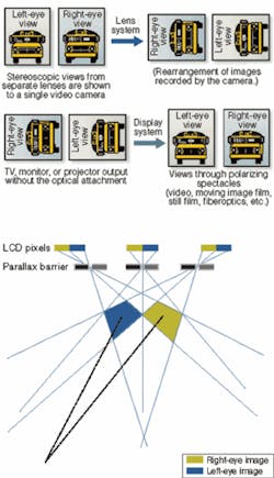
To overcome these problems, SIS has developed and patented an optical-based system that is elegant in its simplicity. To obtain a stereo image, two views from separate lenses are imaged onto a single CCD or CMOS camera by optically rotating each image. Because the 16:9 aspect ratio of both images is rotated 90° counterclockwise, both images fit the 4:3 aspect ratio of the image sensor (see figure). “Regardless of whether the camera is progressive-scan or interlaced, every line captures data about both left and right eye images. Because each image is updated only one half a line from its partner, rather than field or frame sequentially, the result is a flicker-free image without the addition of memory or a modified frame rate,” says Christian.
At this year’s IPOT, held in Birmingham, UK, SIS demonstrated its Add-Depth optical system in use with a USB camera from Videology Imaging Solutions (Greenville, RI, USA; www.videologyinc.com). The advantage of using a single USB camera is particularly important when trying to process the depth information. With a single data feed, processing overhead is reduced, as is initial calibration and setup.
Add-Depth USB sensors are offered in two versions with interocular distances of 45 and 63 mm. Both left and right images are captured on the camera’s CCD imager, simultaneously locked together, and automatically synchronized for display. At present, SIS offers both NTSC and PAL versions of the camera, both with USB digital output. Standard analog video versions are also available.
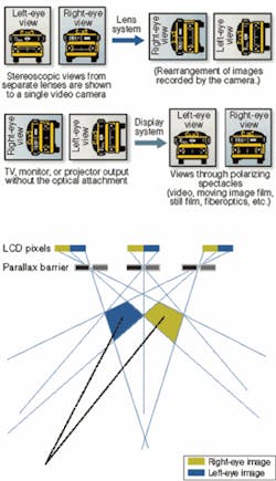
To view images on autostereoscopic displays, SIS offers PC-based software that allows images to be captured, reoriented, and displayed in stereo or as two separate images. At IPOT, SIS demonstrated the camera using an Actius RD3D autostereoscopic 3-D notebook from Sharp Systems of America (Huntington Beach, CA, USA; www.sharpsystems.com). Equipped with a TFT 3-D LCD, the display achieves a 3-D effect using a parallax barrier technique to separate left and right images (see figure).
This parallax barrier is a series of vertical slots that are designed to focus pixels in different directions. Thus, two images are displayed concurrently. By ensuring the “Viewing Diamonds” are set at the same separation as the human eyes (typically 2 1/2 in.), image separation is performed resulting in a stereoscopic 3-D image conveyed from the chosen camera position.
Already, a number of customers have adopted SIS technology for specialized applications. The primary market has been in the tele-operation of unmanned ground vehicles, according to Christian. One medical endoscope manufacturer has also integrated the optics into a range of 3-D endoscopes for industrial applications and a laparoscope for medical research. The scopes produce true stereoscopic images down to a distance of 4 mm.
Although autostereoscopic displays and wireless linked hand-held binocular viewers are compatible, medical applications requires captured images to be viewed using polarized glasses on a SIS 3-D rear-projection system requiring only one projector. This allows freedom of movement sideways and simultaneous viewing by surgeons of differing stature. Captured images can then be analyzed with SIS PC-based measurement software.

