Digital imaging enhances ophthalmic examinations with retinal details
Digital imaging enhances ophthalmic examinations with retinal details
By Michael McMinn
Until recently, Dr. Scott Grant, a Fullerton, CA-based ophthalmologist specializing in retinal and vitreous diseases, had envisioned digital imaging technology as an innovative process but still not good enough for producing human-eye images. He continued to rely on film cameras to record retinal images of his patients. However, after he performed an updated analysis, Dr. Grant acknowledged, "Digital cameras have reached a point where they have practical [retinal] diagnostic applications. I decided the time was right to go digital."
Current high-resolution digital cameras not only give ophthalmologists the ability to instantly see the back of a patient`s eye, but provide those images in greater detail than previously obtained. Rather than wait a day or more for conventional film processing, physicians can now diagnose a patient immediately.
For both physician and patient, instant eye images convey several advantages. Digital images result in better patient medical care because problems, such as bleeding in the back of the eye (known as leakage), can be treated promptly. The patient also avoids a second appointment to discuss the results or receive treatment.
For the physician, the ability to examine, diagnose, and discuss treatment for a patient during the same appointment is time- and cost-efficient. Depending on the circumstances, treatment can be done immediately after the diagnosis. Eliminating a second visit translates into decreased costs.
Some existing video and low-resolution digital imaging systems do not possess the resolution or image depth to capture the required eye details for challenging diagnoses. In the case of bleeding, for example, the ophthalmologist may be able to see the leakage, but the images may not reveal the source or other critical details. Moreover, if an eye has an increased level of opacity, some details may not even be seen. In the past, expensive film and processing would have been needed to identify such details. Now, the latest high-resolution digital imaging technology provides increased levels of detail that overcome most eye-imaging obstacles.
Imaging integration
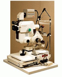
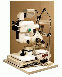
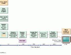
Zedec Technologies (Durham, NC), an integrator of machine-vision and medical-imaging systems, has developed a novel retinal-imaging system using the Digital Equipment Corp. (Maynard, MA) Alpha processor and Eastman Kodak (Rochester, NY) MegaPlus camera, model 1.6i (see Fig. 1). When an ophthalmologist triggers the digital camera, a 2-Mbyte image is compressed, autocontrasted, stored, and displayed in less than 750 ms (see Fig. 2).
The Kodak digital camera is a 10-bit charge-coupled-device (CCD) camera. Many CCD cameras are 8-bit types, which provide 256 levels of gray. The Zedec system provides 1024 levels of gray. The higher image level allows ophthalmologists to see individual blood vessels and the source of bleeding that would otherwise shroud those vessels. With only 256 levels of gray, the ophthalmologist can adjust the system contrast and brightness but is not going to see details. With a 10-bit camera, the ophthalmologist can see which blood vessels are feeding the leak, which is a critical attribute.
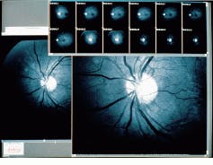
In addition, ophthalmologists, or laboratory technicians, can select a particular group of the available 1024 levels of gray they want to view. So, instead of having to adjust the image contrast or brightness, the physician can select a range of image information to view the retina more clearly (see Fig. 3).
Zedec demonstrated the imaging system, which runs under Windows NT software, to Kowa Optimed Inc. (Torrance, CA) using Kowa`s fx-500 retinal camera. Soon after, the two companies signed a joint venture agreement. Kowa Optimed distributes several medical imaging and optic devices that are manufactured by its parent company Kowa Electronics and Optics (Tokyo, Japan). Kowa`s fx-500 and fx-500S precision Fundus cameras provide color, fluorescein, video, and Polaroid imagery. These retinal cameras offer high-resolution digital imaging capabilities as well.
The digital imaging portion of the complete retinal system is called Komit (Kowa Ophthalmic Medical Imaging Technology). The system includes a compact-disk (CD) recorder so ophthalmologists can record patient data and images to CD-ROMs for permanent storage. A database on the hard drive keeps track of all the patients` information.
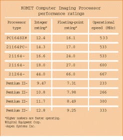
The Komit system computer is generally a Windows NT-based workstation that incorporates either an Alpha processor from Digital Equipment Corp. or a Pentium II processor from Intel Corp. (see table on p. 29). The Alpha processor is typically run at 533 to 667 MHz; the Pentium II typically runs from 233 to 333 MHz. Many integer-type digital imaging operations are better served by the Pentium processor. The more compute-intensive functions, such as image merge, overlay, interpolated zoom, and cine display with alignment, require the floating-point capabilities of an Alpha processor. The system processor design choice depends upon the ophthalmologist`s particular application requirements.
System software
Zedec Technologies has developed a custom software toolkit that includes an image-processing library, an image-measurement library, an Operating System Independent (OSI) library, a Graphical User Interface Independent (GUII) library, and hardware-specific libraries. These libraries allow system development across operating systems such as Windows NT, Windows 95, Digital UNIX, and Linux. The KOMIT system for Kowa, as well as the Zedec menu-driven quantitative imaging software package (QuantIm), uses these libraries.
In addition, company memberships in developers association programs of both Microsoft (MS Developer Network) and Digital Equipment (Association of Software & Application Partners) have proven valuable. Through these associations, the digital imaging toolkit is kept updated and ready for the next version of the respective operating systems. Currently, new ophthalmic software under Windows NT 5.0 and Windows 98 is being tested.
To develop the ophthalmic system for Kowa, Zedec interviewed users in the industry to determine the system requirements. From these requirements, the proper hardware was selected to do the job, such as the Kodak digital camera and the Bitflow Data Raptor camera interface. The DEC Alpha-based workstation was chosen because of its fast speed.
Next, system requirements dictated a challenging one image per second for each acquisition. With a 10-bit camera at 1280 x 1024 pixels, the images needed 2.5 Mbytes, a large amount of data to process in one second. Existing ophthalmic systems use 8-bit cameras at 1024 x 1024 pixels and deliver 1 Mbyte/image. The developed system`s PCI interface, high-speed components, and fast Alpha processor can expose and read the CCD, store to disk, create a thumbnail, autocontrast, and display a thumbnail in 750 ms, thereby achieving the 1-s/image requirement.
Another critical design issue was the user interface. Because most ophthalmic technicians are not computer experts, the user interface had to be easy to use. Because the many layers of menus typically used in most software products were not appropriate, the developed system incorporates quick access buttons that initiate the fundamental system procedures used most often. The more esoteric functions are accessed from typical menus. This button-style access is possible through the use of the custom imaging-software libraries. The final design result is a fast, easy-to-use system at a competitive price.
Fast digital images
"Until now, digital imaging was always used with the caveat that it wasn`t as good as film," says Craig Ross, vice president, marketing and sales, Kowa Optimed. "The [digital] camera changes all of that. Its resolution closely approaches that of film. And with the Alpha processor, digital images are handled as fast as the camera can shoot. While digital imaging had already been accepted as a usable tool, this system removes all reasons for not going digital."
For Dr. Grant, the speed and quality of a KOMIT system convinced him that now was the time to go digital. "This digital imaging system is technologically superior to other systems I`ve seen," he says. "While film is still superior in terms of resolution, the digital system is diagnostically accurate, and it has the advantages of instantaneous information and the elimination of the darkroom," he adds. The Kowa camera does allow for simultaneous film and digital imaging.
The vast majority of retinal imaging is done via fluorescein angiography. A fluorescein dye is injected into a patient`s arm through an intravenous connection. Using special filters on the retinal camera, the ophthalmologist can record how the blood is flowing through the back of the eye. This process reveals problems associated with the retina and other structures in the back of the eye.
"The ophthalmologist can see the dye come through the blood vessels," Ross explains. "This makes diagnosis far superior to a simple color image. With a color image, sometimes it`s difficult to see the source of the bleeding. And if you get a red cloud in the retina, it becomes almost impossible to see where the bleeding is coming from and where it`s ending up. The dark areas show where there isn`t any blood flow," he notes.
The need for processing speed comes from how fast the images are taken. Approximately 36 images are taken in less than two minutes for both eyes. Images are taken in rapid succession to adequately document the flow of flourescein.
"Because the image can be viewed immediately on the monitor, I can quickly adjust the camera-flash intensity if necessary," states Kathy Carpenter, Dr. Grant`s photographer. "Any images that prove to be nonuseful because of any type of artifact can be removed."
Using the digital imaging system is almost identical to using film except there is no camera to load nor any film to process. The technician acquires a "red-free" image of the retina. This "red-free" image is taken by rotating a green filter in the image path. This image is used as a reference for the retina structures. Several images are taken of both eyes.
After a fluorescein intravenous injection (typically in the arm) is administered to the patient, the technician also starts an image timer. Typically within 5 to 10 s, the technician begins taking early images of the fluorescein as it enters the eye through the arterial system.
The technician continues to take images through the "mid-phase" of the fluorescein flow through the bloodstream. Within 20 to 40 s, the flourescein has circulated and is showing in the venous system. The technician usually pauses for two to five minutes and then takes more images to visualize any "pooling" or "leaking" in the membrane.
At this point, Dr. Grant evaluates the images to determine what treatment is needed. By displaying the digital images on a monitor, he can enlarge and enhance the images to bring out details that were not available before. This capability is useful when, for example, a cataract is present. A cataract will blur the image and reduce the contrast. Digital enhancement can often be used to obtain views of details that have been obscured by the cataract.
"Now we can go in and use the enhancing capabilities to adjust the image to bring out that detail," Ross says. "That`s important when it comes to ICGs (indo-cyanine greens), which is another type of injected dye. It`s a cyan-green dye that is used to see different structures of the back of the eye. Those images tend to come out dark, so being able to move along the 1024-gray-scale level to find the image and bring out the detail are important attributes."
If the problem is leakage, laser treatment is usually the method used to stop it. After the treatment, the fluorescein procedure is repeated, and the two images sets are compared to determine the treatment`s effectiveness. Says Dr. Grant, "It is valuable to explain the medical problems immediately without having patients wait and worry. We can arrange for treatment expeditiously."
Digital imagery also dropped the cost of materials associated with retinal tests at Dr. Grant`s office by about one-third because film, processing, and paper are no longer used. Because the images are kept digitally on CD-ROM, large storage equipment and space are also not needed. "In three years we expect to recapture the cost of the system due to the savings in film processing costs and in darkroom time," claims Dr. Grant.
Furthermore, digital imagery also has the advantage of being able to transmit imaging files over a network using a modem to a central office for review or to another ophthalmologist for a second opinion. Moreover, the doctor can be in one office and view images taken at other locations, thereby providing near-real-time examination.
"The bottom line is that digital imaging provides instantaneous information that can immediately be used to diagnose an eye problem and allow for immediate treatment," Grant says. "This is a great convenience, and it significantly improves patient care."
MICHAEL MCMINN is president of Zedec Technologies, Durham, NC 27713.
FIGURE 1. The Kowa fx-500 professional Fundus camera incorporates Zedec Technologies`digital retinal system into its Kowa Ophthalmic Medical Imaging Technology (KOMIT) system. This digital system generally uses a DEC Alpha processor to handle the large digital files captured by the Kodak MegaPlus camera, model 1.6i. Along with its PCI interface, the system can expose, read the CCD camera, store to disk, create an image, perform autocontrasting, and display a final image in 750 ms.
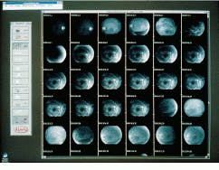
FIGURE 2. In the Kowa Ophthalmic Medical Imaging Technology (KOMIT) retinal-eye imaging system, the user is in full system control via custom system software. System operation is based on user-selected functions, for example, "Start Acquire." The system functions are repeated until all the retinal images needed are obtained; as many as 144 images can be obtained for each patient record. One system operational image sequence takes 750 ms.
FIGURE 3. In the retinal imaging system, a Kodak MegaPlus Model 1.6i camera uses a 10-bit sensor and 1024 levels of gray scale that allow ophthalmologists to quickly acquire, enhance, and enlarge eye-retina images. An off-the-shelf 21-in. color monitor displays a stream of images (right), any one of which can be enlarged. In a healthy human eye (below), the optic nerve is surrounded by blood vessels in the center. The enhanced image information enables the digital viewing, processing, and display of increased retina details for improved diagnosis.
Aspen Systems Inc.
Wheat Ridge, CO 80033-3865
(303) 431-4606
Fax: (303) 431-7196
Web: www.aspsys.com
Bitflow Inc.
Woburn, MA 01801
(781) 932-2900
Fax: (781) 933-9965
E-mail: [email protected]
Web: www.bitflow.com
Digital Equipment Corp.
Maynard, MA 01754
(978) 493-5111
Fax: (978) 493-7374
Web: www.digital.com
Eastman Kodak Co.
Rochester, NY 14650
(716) 722-4385
Fax: (716) 477-4947
Web: www.kodak.com/go/ccd
Intel Corp.
Santa Clara, CA 95054
(408) 765-8080
Fax: (408) 765-1402
Web: www.intel.com
Kowa Optimed
Torrance, CA 90502
(310) 327-1913
Fax: (310) 327-4177
E-mail: [email protected]
