Digital x-ray system puts pets in perspective
Andrew Wilson, Editor, [email protected]
American families spend almost $10 billion a year on veterinary care services for their pets. For manufacturers of medical imaging equipment, the move from film-based x-ray to digital x-ray systems provides an opportunity to eliminate hazardous and expensive chemical-based film-based processing while, at the same time, reducing the costs of the systems.
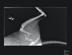
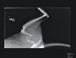
By providing digital images with a single exposure, images can be enlarged and manipulated, displayed to clients on any networked monitor in the clinic, e-mailed to specialists for telemedicine consults, and stored as a computer file. Better still, the wider latitude allowed by digital capture and the nearly instant display of the image on the monitor while the animal is still on the table eliminates unnecessary radiation and stress to the pet and the operator while eliminating the space required to store radiographic film.
Unlike x-ray systems designed for human beings, however, those designed for veterinary applications are less expensive and often must be retrofitted to existing examination tables. Due to the small size of the patient, spatial resolution must be greater than for human systems. For this reason, RemoteVet (Kennesaw, GA, USA; www.remotevet.com) has developed the Merlin D-Rad Retro-fit system, an imaging subsystem that allows existing analog veterinary examination tables to be converted to digital imaging workstations in approximately three hours. Using a clinic’s existing x-ray generator, retrofitting costs and equipment training are reduced.
“In the design of existing film-based systems,” says David Gardner, president of Salvador Imaging (Colorado Springs, CO, USA; www.salvadorimaging.com), “a pet is placed on a 17 × 17-in. film plate that is exposed using an x-ray generator. The film is developed and the x-ray viewed on a light console by the physician.” To retrofit these systems, this film plate is replaced with a gadolinium oxide scintillation plate. When subjected to x-ray energy, this plate emits faint green light that must be captured by a digital camera.
“However,” says Gardner, “exposing CCD-based cameras directly within the x-ray path can result in ionization of the silicon dioxide atoms in the layer separating the gate structure from the buried channel in the CCD. This charge is trapped in the dielectric, accumulates over time, and affects the charge-transfer efficiency of the CCD.” Because of this, the Merlin D-Rad system incorporates a 45° mirror that is placed directly under the scintillation plate. “In this way,” says Gardner, “a CCD camera can be mounted perpendicular to the x-ray path and thus can reduce any radiation effects that may occur.”
In the design of RemoteVet’s system, an SI-6M1-FF camera from Salvador Imaging (Colorado Springs, CO, USA; www.salvadorimaging.com) captures reflected images from the scintillation plate. Using a KAF-6303E full-frame image sensor from Kodak Image Sensor Solutions (Rochester, NY, USA; www.kodak.com), the camera is capable of digitizing images at 3072 × 2048 ×12-bit resolution. Captured images are then transferred to a PC-based X64-CL frame grabber from DALSA (Waterloo, ON, Canada; www.dalsa.com).
Once images have been captured by the system they can be stored on the system’s 250-Gbyte hard drive or displayed on a high-resolution flat-panel monitor. To perform image-processing functions, RemoteVet developed a full suite of image processing, enhancement, archival, and networking software specifically tailored to veterinary x-ray images. This package allows the operator to perform on-screen measurements and allows technicians to magnify images, adjust for bone vs. soft tissue exposures, and alter resolution and image-contrast settings. Because these images are DICOM-compliant, they can also be networked within the clinic or to remote workstations for further diagnostic analysis.

