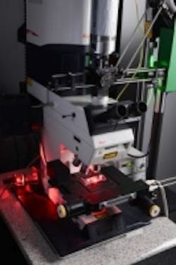
Fraunhofer imaging technique enables faster testing of drug ingredients
Researchers at the Fraunhofer Institute for Production Technology have combined two microscopy techniques that reduces the time required for testing of new drug ingredients on biological cells by anywhere from 50 to 80%.
During the process of developing new medications, biologist and pharmacologists test different active ingredients and chemical compounds, with the goal of finding out how biological cells react to the substances, according to the Fraunhofer Institute. Researchers often use a fluorescence microscope that produces holographic images so the cells can be viewed in 3D. This is done by creating an optical image and digitizing it for recording and analysis on a computer, which then calculates the data needed to display the 3D image of the cells. This method enabled the researchers to examine the cells without touching them and without having to use markers to make the cells visible.
Previous techniques of obtaining precise and reliable answers of how cells react to new chemical substances required a great deal of time and energy for scientists, but combining the technique of digital holographic microscopy with the use of optical tweezers cuts a significant portion of time off the procedure. Optical tweezers are a special instrument that uses the force of a focused laser beam to trap and move microscopic objects, which enables the researchers to pick up selected cells and transfer them to individual wells of a microarray for testing, and keep them trapped there.
"By combining these two instruments, we can save between 50 and 80 percent of the time normally needed for such work, depending on the type of cell and the test method employed. That’s mainly because we don’t have to carry out so many repeated measurements," explains IPT group manager Stephan Stürwald in the Fraunhofer press release.
The system is very easy to use, and the optical tweezers can also be controlled via the touchscreen of a tablet PC. It will also enable the researchers to use the laser in the system to selectively destroy cells that are unsuitable for testing. A system prototype was developed consisting of the two modules, and the researchers were able to carry out initial tests using it.
View the Fraunhofer Institute press release.
Also check out:
Optical sensor tracks zinc in cells for cancer research
MIT and Massachusetts General Hospital developing new X-ray imaging technology
(Slideshow) 10 different ways 3D imaging techniques are being used
Share your vision-related news by contacting James Carroll, Senior Web Editor, Vision Systems Design
To receive news like this in your inbox, click here.
Join our LinkedIn group | Like us on Facebook | Follow us on Twitter | Check us out on Google +
About the Author

James Carroll
Former VSD Editor James Carroll joined the team 2013. Carroll covered machine vision and imaging from numerous angles, including application stories, industry news, market updates, and new products. In addition to writing and editing articles, Carroll managed the Innovators Awards program and webcasts.
