To determine the most appropriate treatment for diseases such as cancer, researchers must study individual cells and tissues. Their analysis relies on hundreds of immunohistochemical (tissue-based) and immunocytochemical (cell-based) tests, which involve staining samples with commercially available reagents and observing the sample's chemical reaction under a microscope.
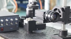
Several automated imaging systems for cellular analysis are currently available for specific pathological and drug-discovery assays; however, the PC host and microscope combination system, Automated Cellular Imaging System (ACIS), from ChromaVision Medical Systems (San Juan Capistrano, CA; www.chomomavision.com) provides a single platform for the evaluations. The ACIS uses a custom-made, bright-field microscope that can accommodate any standard microscope objective as a result of an automated motorized light condenser controlled by a proprietary light-controller board in the PC host. The condenser adjusts the light path and levels based on the needs of the specific assay. This versatility is critical to ACIS operation because, after a tray of 100 slides is loaded into a robotic conveyor system, the ACIS takes a series of images with progressively higher magnification to automatically identify cells that exhibit certain user-set characteristics such as color, intensity, and shape.
Doctor in the software
The ACIS automatically reads a barcode sticker on each slide containing an individual tracking number for the slide, as well as the required procedure. The system takes the first image of the slide under examination with a Sony (Park Ridge, NJ, USA; bssc.sel.sony.com/Professional/) DXC9000 three-chip progressive-scan color CCD camera and sends the image to a PC host through a Matrox (Dorval, QC, Canada) Meteor II/Multi-Channel frame grabber. The image is evaluated on the image-processing board using several primitives from the Matrox Imaging Library, including blob analysis, pattern-matching, and measurement.
"For us to create an image-processing library ourselves would be ridiculous. If you need a function, you reach in the tool bag and 99.9% of the time it's already been implemented. I can worry about the algorithms I need to design and the tools I need to meet the end goal," explains ChromaVision software engineer Jeff Caron.
The ACIS software resident in the PC RAM takes over and converts intensity and color patterns from the images into criteria that can automatically identify suspicious images or regions for later review by a physician. The software can also suggest specific treatments based on what it finds in the images.
The ACIS continues by taking additional pictures of specific regions of interest as necessary at progressively higher magnifications and displays the images in a montage pattern on the PC monitor through a Matrox 550 video card. The images can also be saved to local hard drives or remote server-based SQL databases.
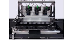
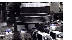
After a slide has been fully evaluated, the ChromaVision conveyor system removes the old slide and places it in the outfeed container before loading a new slide for analysis. The barcode slide information is read, and the process repeats for up to 100 slides per tray.


