X-ray and 3D imaging techniques used to non-destructively visualize ancient fossils
Researchers at theUniversity of Bonn have combined x-ray imaging, 3D imaging, and computer animation in a study to demonstrate the ability to visualize ancient fossils without destroying the sample.
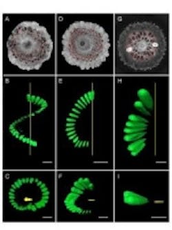
In stark contrast to traditional techniques such as thin-sectioning, which requires the actual slicing of a fossil into thin sections, Dr. Carole Gee and her colleagues have developed a technique that utilizes high-resolution x-ray computed tomography, or MicroCT, to provide virtual cross-sections of a specimen. In aresearch paper, the process for imaging the internal anatomy of 150 million-year-old conifer seed cones was described.
MicroCT was used to image the seeds using aGE Phoenix v|tome|x system for 2D x-ray inspection and 3D computed tomography. These projections were then processed with GE Phoenix datos|x 2.0 CT software and VG Studio Max software, which is integrated with the datos software, to produce image stacks of serial single sections for 2D visualization, 3D reconstructions with targeted structures in color, and computer animations.
In addition to the fact that this form of visualization is non-destructive, Gee also notes that it enables the observation of internal features such as seeds, vascular tissue, and cone scales. Also, by adding artificial color to highlight certain structures or tissues, the natural pattern of growth is observable.
"It’s amazing to visualize internal structures of dinosaur-aged fossils in such great detail without cutting up the fossil or damaging them at all," Gee said in the press release.
As a result of using this new form of imaging, Gee and her team were able to identify the fossils are belonging to three distance families: Pinaceae—the pine family, Araucariaceae—a family of coniferous trees currently found only in the Southern Hemisphere, and Cheirolepidiaceae—a now-extinct family of conifers known only from the Mesozoic.
"This tells us that 150 million years ago, the ancient forests of western North America consisted of members of these three families. The fossil cones of the Araucariaceae from Utah confirm that this family, which now grows naturally in Australasia and South America, once had a worldwide distribution," she said.
Going forward, Gee said she hopes the study will provide researchers with an alternative to traditional techniques and that ultimately, the technique will become commonplace in paleobotany and botany.
View theresearch paper.
View the press release.
Also check out:
3D scans show entire fossil of baby dinosaur skeleton
(Slideshow) 10 innovative current and future robotic applications
Clinical study shows 3D holographic images of the heart during procedures
Share your vision-related news by contactingJames Carroll, Senior Web Editor, Vision Systems Design
To receive news like this in your inbox,click here.
Join ourLinkedIn group | Like us on Facebook | Follow us on Twitter| Check us out on Google +
About the Author

James Carroll
Former VSD Editor James Carroll joined the team 2013. Carroll covered machine vision and imaging from numerous angles, including application stories, industry news, market updates, and new products. In addition to writing and editing articles, Carroll managed the Innovators Awards program and webcasts.
