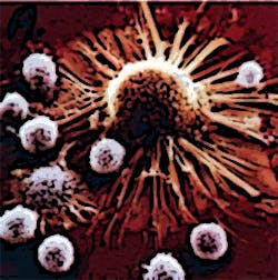
Luminescent bacteria help monitor tumors
One of the biggest problems in the development of cancer therapies has been the targeting of tumors for treatment while leaving healthy, normal cells untouched.
Bacteria have a natural ability to grow inside tumors, a fact that has been known for some time. In fact, when non-disease causing bacteria are injected intravenously, they are attracted to tumors, but fail to gain a foothold anywhere else in the body.
Now, a research team led by Dr. Mark Tangney at the Cork Cancer Research Centre, (CCRC) at University College Cork (UCC; Cork, Ireland), believe that they have taken a key step to discover why and how the bacteria prefer the company of tumors to healthy tissue.
Dr. Tangney and his team have developed a way to track bacteria within tumors in real time to learn more about what makes the tumor environment so attractive to these microbes. By engineering probiotic bacteria to produce luminescent light, the team were able to monitor bacterial growth in tumors as it happened using 3-D bioluminescence imaging.
This scanning method revealed information about the number and location of the bacteria, precisely revealing where the bacteria were living in the tumor.
Imaging the light from bacteria could be combined with CT scanning to provide details of their relationship with different parts of the tumor, such as the blood supply.
The work was performed by Dr. Michelle Cronin, Dr. Sara Collins and others from the CCRC in collaboration with a US industrial partner Perkin Elmer (Waltham MA, USA), a research team at the University of California Los Angeles and researchers at UCC's department of microbiology.
Related articles from Vision Systems Design that you might also be interested in.
1. Spectroscopy aids brain tumor detection
UK researchers have shown that infrared and Raman spectroscopy - coupled with statistical analysis - can be used to differentiate between normal brain tissue and different tumor types.
2. Image processing program detects tumor cells
A researcher at the University of Twente's MIRA Research Institute (Enschede, The Netherlands) has developed image processing software that can count the circulating tumor cells in a blood sample and identify them by type.
3. Imaging helps team to detect cancerous cells
Scientists from The Scripps Research Institute (La Jolla, CA, USA) have demonstrated the effectiveness of a blood test for detecting and analyzing circulating tumor cells (CTCs) -- breakaway cells from patients' solid tumors -- from cancer patients.
-- Dave Wilson, Senior Editor, Vision Systems Design
