High-speed imaging system assists in heart rate variability research
Variations in time intervals between consecutive heart beats known as heart rate variability (HRV) serve as a diagnostic and prognostic tool for cardiac as well as non-cardiac diseases such as heart failure, aging, Parkinson's, diabetes, and sepsis. Typical investigation methods involve determining time intervals from electrode measurements, but a team of researchers from the Medical University of Graz (Graz, Austria; www.medunigraz.at/en) developed a reliable recording method using a high-speed camera and video recording system from which beating rate and contraction strength can be assessed.
For the study, an experimental chamber containing intact sinoatrial node (SAN) mouse tissue was mounted on the stage of a BX51WI Olympus (Waltham, MA, USA: www.olympus-lifescience.com) upright microscope with tissue superfused with oxygenated standard external solution kept at a constant temperature of 23°C. Recordings began 20 minutes after the onset of superfusion—the continuous following of the solution over the outside of the tissue—allowing the tissue to establish and maintain a stable beating rhythm.
An EoSens CL camera from Mikrotron (Unterschleissheim, Germany; www.mikrotron.de/en) captured tissue sample video at 1280 x 1024 pixels. The camera features a 1.3 MPixel LUPA1300-2 CMOS image sensor from ON Semiconductor (Phoenix, AZ, USA; www.onsemi.com), Camera Link interface, frame rates up to 506 fps, and dynamic range up to 90 dB.
The researchers paired the camera with Mikrotron’s MotionBlitz LTR Long Time Recording System, which implements a hardware recording unit to avoid jitter effects of software trigger events, according to the team. After recording the first video, the researchers added acetylcholine to the solution, and after a five minute superfusion, the system captured the second video. Known as a predominant transmitter of the parasympathetic nervous system, acetylcholine’s effects on atrial tissue are well investigated and include a decrease in beating rage and force of contraction, according to an academic paper on the research (http://bit.ly/VSD-BEAT).
"In this study, we utilized video recordings to investigate the variability of intervals as well as mechanical contraction strengths and relative contraction strengths with nonlinear analyses," says Helmut Ahammer, the Medical University of Graz professor who headed the study. "Additionally, the video setup allowed us simultaneous electrode registrations of extracellular potentials."
Related: TDI cameras solve new challenges in DNA sequencing
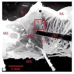
Close to the primary pacemaking site of the tissue, a 160 x 160 region of interest showed distinct contraction, providing maximum grey level changes during the beating of the issue (Figure 1). Larger ROIs yielded overly large video files, while smaller ROIs produced low signal-to-noise ratios. Regions with thick tissue layers moving into the ROI led to the highest signal to noise ratios. With a sample rate of 1,000 fps and a recording duration of 5 min, 300,000 single uncompressed images (160 × 160 resolution, 25,600 pixels) were taken and stored on the MotionBlitz's hard disk in avi format, with one avi file requiring 21 GB of memory.
Despite using a monochrome camera, the system saved the individual images in RGB format. Monochrome cameras typically provide higher signal to noise ratios than color cameras, which is important in high-speed acquisitions with small exposure times. Images were converted to 8-bit, lowering the memory demand to 7.6 GB per video. The researchers tested the setup against electrical and optical inferences coming from ambient light sources such as the laboratory light or the microscope’s light source. Fourier analyses of static scene videos revealed no residual frequency components of power supply frequencies or other additional noise components.
Recordings took place over a period of several hours, providing the team with ample amounts of data for further analysis. Recorded images stored directly on a RAID protected drive array, eliminating download times. Professor Ahammer's team evaluated beat to beat interval and contraction strength variabilities with the system. Each image recorded contractions of the tissue as changes in average grey values. Simultaneous measurements of extracellular potentials using a cardiac-near-field electrode validated beat to beat intervals of the video recordings.
In summary, the Mikrotron video recording technique offers a promising tool for investigating beat to beat behavior regarding absolute values of beating rate and contraction strength, as well as their variabilities in autorhythmic tissue, according to the team.
About the Author

James Carroll
Former VSD Editor James Carroll joined the team 2013. Carroll covered machine vision and imaging from numerous angles, including application stories, industry news, market updates, and new products. In addition to writing and editing articles, Carroll managed the Innovators Awards program and webcasts.
